SEARCH

Stollery N. Sun damage. Practitioner 2015; 259(1784):28-29
Sun damage
05 Aug 2015
AUTHORS
Dr Nigel Stollery MB BS DPD, GPwSI Dermatology, Kibworth, Leicestershire, UK
Article
Actinic keratosis
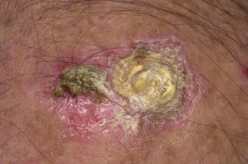
Actinic keratoses are common skin lesions related to sun exposure particularly UVA. They are benign but may be premalignant and are a risk factor for squamous cell carcinoma (SCC). In individuals with ten or more lesions there is a 15% incidence of progression to SCC over five years (most commonly on the hands, wrists and forearms). Actinic keratoses usually occur on sun-exposed areas of the head, hands and neck but can also develop on the lips and rarely on the conjunctiva. In patients with vitiligo actinic keratoses may develop in the depigmented areas. Treatment includes cryotherapy, topical preparations such as 5 fluorouracil and surgery.
Photodermatitis
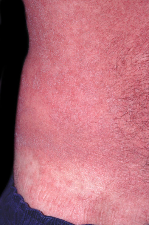
Photodermatitis is a type of allergic contact dermatitis in which the allergen is activated by UV light leading to an allergic response. Further exposure to light then produces a photo allergic rash which is usually like eczema. Often the skin will swell with an associated burning feeling followed by the development of small vesicles. The skin is also usually very itchy as the rash develops and may be urticated. After this has settled post-inflammatory hyperpigmentation may persist especially on the face. A wide range of drugs can cause photodermatitis including amiodarone, retinoids, some NSAIDs, St John’s Wort and benzoyl peroxide. When these drugs are used patients need to protect their skin using high factor sunscreens (SPF 30 and above) and ideally cover up, or avoid going out, in the sun. In rare cases, where patients are very sensitive to the sun, azathioprine or short-term oral steroids can be helpful.
Polymorphic light eruption
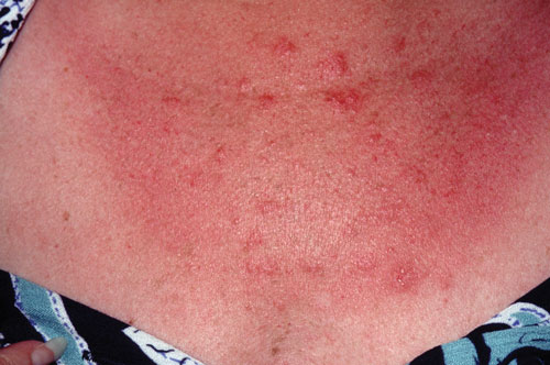
There are many different light-induced conditions that affect the skin and differentiating between them can be difficult. There are often subtle differences which can be determined with a very careful history. These include factors such as the distribution of the rash and the time it takes for it to occur after exposure to UV light. Polymorphic light eruption is one of the most common light-induced conditions. It usually occurs hours after exposure with non-exposed areas spared from the rash. Between 10 and 15% of the UK population are thought to be affected by one of the two types of rash. Most individuals develop multiple papules 2-5 mm in diameter, while others develop blisters which coalesce into much larger areas resembling eczema. There is no treatment for polymorphic light eruption but avoiding UV light, and the use of sunscreens, can be very helpful.
Sunburn
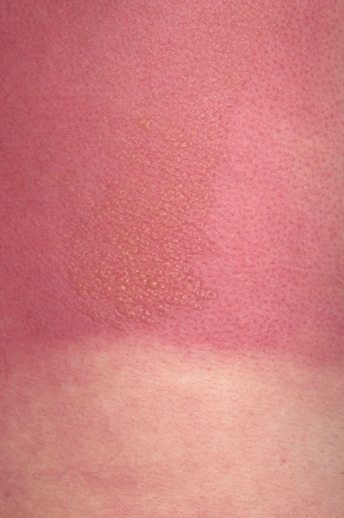
On exposure to UV light two forms of tanning occur. Immediate pigment darkening develops after exposure to UVA as melanin is released by melanocytes. This begins to fade within a few hours. The second is delayed tanning which takes about three days to develop and lasts several weeks. Exposure to high-dose UV radiation will also start to damage skin cells. The degree of damage varies with skin type, UV intensity and exposure. When burning occurs skin cells in the upper layers are destroyed and shed leaving the newer cells below exposed to further UV damage. Acute burning causes inflammatory changes, with the release of inflammatory mediators, and vascular changes which affect cell proteins and DNA. This sequence of events, especially if occurring repeatedly, may lead to malignant transformation and the development of tumours such as melanomas, and squamous cell and basal cell carcinomas.
Solar lentigenes
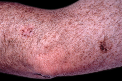
More commonly known as age spots or liver spots, solar lentigenes are small pigmented macules with a clearly defined edge and normal adjacent skin. They occur with chronic UV light exposure and usually evolve over many years, although rarely they may appear very suddenly. They can occur anywhere on the body but are more common on the face, arms and hands. When viewed under the microscope affected areas have increased numbers of normal melanocytes in the basal layer of the epidermis. Although they are benign their presence indicates chronic exposure to UV light which increases the risk of melanoma. Careful examination is therefore advised to make sure melanomas are not missed. This patient also has a benign ink spot lentigo which appears as a dark spot on a background of multiple lentigenes (on the right of the picture).
Malignant melanoma
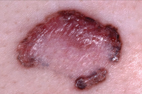
The incidence of melanoma has increased significantly over the past decade, however, it remains a relatively uncommon type of skin cancer. NICE recommends that patients with a suspicious lesion that may be a melanoma are referred urgently, using a suspected cancer pathway referral, for an appointment with a specialist within two weeks (www.nice.org.uk/guidance/ng12). Surgical excision is the mainstay of early treatment. In recent years the management of advanced melanoma has changed with the development of novel drugs such as biological therapies. NICE has just published a clinical guideline on the assessment and management of melanoma (www.nice.org.uk/guidance/ng14).
ARTICLE IN PDF
 Download this article in PDF format