SEARCH

Stollery N. Scalp conditions. Practitioner 2015; 259(1778):32-33
Scalp conditions
22 Jan 2015
AUTHORS
Dr Nigel Stollery MB BS DPD, GPwSI Dermatology, Kibworth, Leicestershire, UK
Article
Squamous cell carcinoma
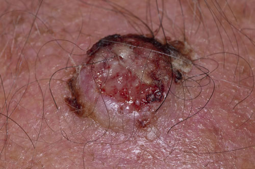
Squamous cell carcinoma (SCC) is a potentially fatal form of skin cancer that can occur anywhere on the body. As UVR exposure is a significant risk factor, the balding male scalp and ears are high-risk areas. UVR induces DNA damage leading to keratinocyte transformation and an alteration of the cutaneous immune response which increases the risk of SCC. Immunosuppression, HPV virus infection and exposure to chemical carcinogens can also increase the risk of SCC. Signs of SCC development include crusting, ulceration, bleeding and in some cases the production of excess keratin in the form of a cutaneous horn. If there is diagnostic doubt a biopsy can be performed. The treatment of choice is excision which may be by Mohs micrographic surgery. In cases where surgery is not possible radiotherapy is an alternative option.
Intradermal naevus
Intradermal naevi are very common benign moles that usually develop from late childhood onwards, but rarely after the age of 60. Although they do contain melanin, this is located deep in the dermis rather than at the dermoepidermal junction, hence the skin-coloured appearance of the lesions. They vary in size from 5 mm to 1 cm in diameter and tend to be raised or pedunculated. Intradermal naevi are usually diagnosed by their clinical appearance and treatment is not generally required. In this case, the size and location of the naevus meant it was regularly traumatised by combing the hair and so it was removed by a simple shave excision.
Actinic keratosis
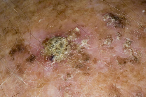
Actinic keratoses are the most common skin lesions with the potential for malignant transformation into squamous cell carcinoma. They arise on sun-exposed areas in response to UV light. Typically they occur as areas of rough palpable skin which may be red and scaly and increase in size with time. They are usually 3-10 mm in diameter but in some cases larger lesions measuring several centimetres across may develop. Rarer variants include brown pigmented, atrophic, bowenoid, Lichen planus-like and hyperkeratotic (horn-like) actinic keratoses. If the diagnosis is in doubt a biopsy can be carried out to exclude squamous cell carcinoma. Treatment can be divided into surgery and topical therapies which include cryotherapy, 5-fluorouracil, imiquimod, diclofenac gel and ingenol mebutate gel.
Seborrhoeic keratosis
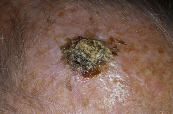
Seborrhoeic keratoses are very common benign skin lesions which occur with increasing frequency with age. Despite their name the sebaceous glands are not involved and the lesions do not occur in sebum-rich areas. Although seborrhoeic keratoses are not associated with UV exposure, they can develop after sunburn or dermatitis. Their variation in colour from white to black means that they are often mistaken for malignant lesions such as melanoma. Dermoscopy can be helpful in making a diagnosis as the lesions have a very characteristic appearance. Very rarely, multiple eruptive seborrhoeic keratoses developing over a short period may be associated with an underlying malignancy, the so-called sign of Leser-Trélat. In the majority of cases treatment is not required but options include cryotherapy, curettage and cautery, shave excision and laser therapy.
Scalp psoriasis
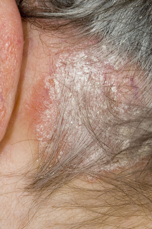
Psoriasis is a chronic inflammatory disease that is associated with hyperproliferation of keratinocytes in the epidermis. It occurs in approximately 2.2% of the population and affects the skin, scalp, nails and joints. Patients with psoriasis have a higher risk of ischaemic heart disease, stroke and peripheral vascular disease and those with severe psoriasis are at greatly increased risk of chronic kidney disease. The PASI (Psoriasis Area and Severity Index) score and DLQI (Dermatology Life Quality Index) can be used to assess patients and guide therapy. The treatment approach depends on the severity and extent of the condition and includes topical therapy, light therapy, and in more severe cases systemic therapy.
Sebaceous naevus
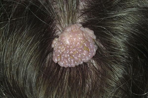
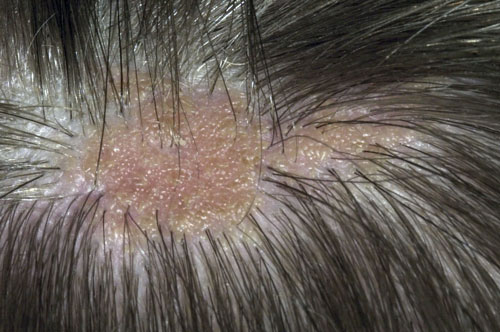
Sebaceous naevi, also known as organoid naevi, are an uncommon type of naevus and are present from birth. They are generally found on the scalp, but can also occur on the face, neck or forehead. Sebaceous naevi are a form of epidermal naevus and involve the epidermis, sebaceous glands, hair follicles, apocrine glands and connective tissue. They are thought to be caused by a mosaic genetic abnormality. Sebaceous naevi tend to increase in size during adolescence and become more wart like. They have a very distinctive appearance by which the diagnosis is usually made. The majority remain the same throughout life and are asymptomatic. Rarely tumours such as basal cell, squamous cell, and sebaceous carcinomas may develop within the naevi. If malignant change is suspected the whole naevus should be excised and sent for histology.
 Download this article in PDF format