Cellulitis
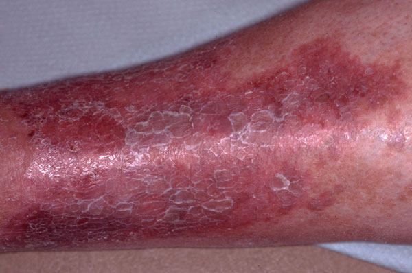
Cellulitis is a non-necrotising inflammation of the skin and subcutaneous tissues which is particularly common in the lower limbs when there is impaired circulation. It is oten caused by acute infection which usually enters the skin through a portal, but this is not always apparent and may only be visible at a microscopic level. The cardinal features are warmth, pain, swelling and erythema. In more severe cases there may also be associated pyrexia and systemic symptoms such as vomiting and malaise. It is unusual to have bilateral infection. The culprit is generally a staphylococcal infection and treatment with an oral antibiotic is required. In very rare cases fungal infections from tinea pedis can cause cellulitis in a limb.
Gastrocnemius atrophy
Focal muscle atrophy may affect any muscle. It has a wide variety of causes including infection, trauma, spinal cord disorders, physical agents, and genetic and enzyme defects. In this case, a prolapsed disc at the L5/S1 level caused damage to the nerve. Around 45-50% of
disc herniations in the back affect the L5/S1 intervertebral disc. Damage to the nerve at this level can also affect ankle plantar flexion and cause sensory loss in the lateral foot and heel. In this patient the injury had occurred a number of years ago leading to permanent atrophy of the muscle despite exercise and physiotherapy.
Infected arthritis of the knee
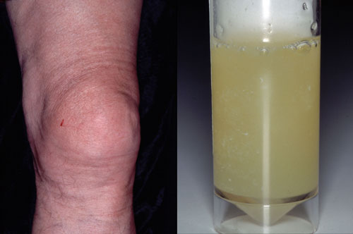
In more than half of cases of septic arthritis it is the knee that is affected. Symptoms include pain, swelling, tenderness and systemic symptoms such as fever. In most cases the causative organism is Staphylococcus aureus which usually enters the joint following trauma or via the bloodstream. Infection can also occur as a complication of surgery including joint replacement and arthroscopy, and is more common in those with rheumatoid arthritis, immunocompromised patients e.g. patients on chemotherapy or those with HIV, and IV drug users. Infected arthritis requires urgent treatment to prevent permanent damage to the affected joint. Treatment includes aspiration of the joint fluids which can then be cultured, IV or oral antibiotics, and splinting of the joint to decrease the pain associated with movement, followed by physiotherapy.
Lymphoedema
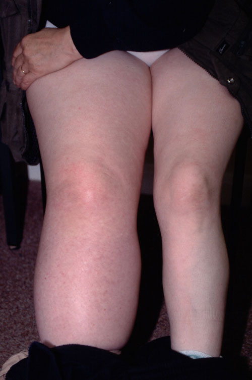
Lymphoedema occurs when lymphatic drainage is obstructed causing protein-rich fluid to collect in the interstitial tissue. This increases fluid retention leading to swelling of the limb. In 80% of cases the lower limbs are affected, but lymphoedema can also occur in the arms, trunk, face and genitalia. Affected areas are more at risk of infections such as cellulitis and ulceration. Diagnosis is usually made on clinical appearance but investigations may be required to determine the underlying cause. Treatment is aimed at increasing function, reducing complications such as infection and trying to improve the appearance of the affected area. It usually consists of physical therapies rather than surgery or medication.
Necrobiosis lipoidica
This is a rare inflammatory skin disorder characterised by irregularly shaped, shiny reddish-brown lesions which develop over a number of months. With time these become yellow, depressed and atrophic and may ulcerate if traumatised. Lesions are most common on the legs but can also occur on the face, arms, scalp and trunk. Although there is a high prevalence of diabetes in patients with necrobiosis the condition can also occur in individuals who do not have diabetes. Necrobiosis lipoidica most commonly presents in the fourth decade but occurs earlier in patients with diabetes. If the diagnosis is in doubt a biopsy can be helpful but healing rates are often poor following a biopsy. There is no effective treatment but it is important to protect affected areas. In painful, inflamed cases topical steroids may help but are less effective once the areas have burnt out and may increase atrophy. Other treatments include intralesional steroids, ciclosporin, topical tacrolimus and anti-TNF therapies.
Allergic contact dermatitis
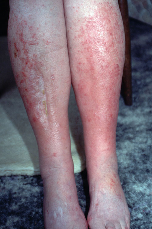
Although rare, allergic contact dermatitis to creams can occur. In this case, a topical steroid cream was responsible for the worsening redness on both lower limbs. With an original diagnosis of varicose eczema the patient had been using emollients along with topical steroids for many months. Initially this seemed to help but after a time the steroid cream caused an increased erythema, pruritus and soreness after it was applied. The patient was referred to a dermatologist and patch testing confirmed sensitivity to the steroid cream which was then stopped and replaced with an ointment.
 Download this article in PDF format