Acanthosis nigricans
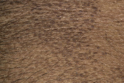
Acanthosis nigricans is an asymptomatic darkening and thickening of the skin, most typically in the axillae, groin and posterior neck. It usually starts as hyperpigmented macules which then progress to palpable plaques. The rash is symmetrical and has a velvety feel. Affected areas may be itchy. Although the condition is usually benign, it may be associated with an underlying malignancy. In 30% of cases the rash appears before any signs of the underlying cancer. Acanthosis nigricans is also associated with other conditions including obesity, lupus, scleroderma, hyperinsulinaemia, acromegaly and dermatomyositis among others. Treating the underlying cause may improve the appearance of the skin. There is no easy treatment approach. Management aims to improve the cosmetic appearance of affected areas and treatment options include dermabrasion, surgical excision and laser therapy.
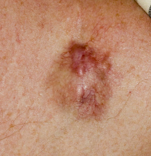
Morphoeic basal cell carcinoma
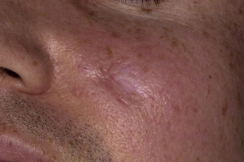
Morphoeic basal cell carcinoma (BCC) is one of the least common types of BCC and probably the most difficult to diagnose clinically.The lesions are mainly found on the face. Unlike nodular BCC, which have rolled edges and ulcerated centres, these lesions often look more like scars with indistinct borders and a yellow waxy surface which is firm to the touch. Telangiectasia is often present which can be a helpful clue, but the mainstay of diagnosis is biopsy and histological examination. The lack of a clear border means that the lesions can be difficult to excise adequately and may recur. The treatment of choice is Mohs micrographic surgery which has the highest cure rate. In this procedure tissue is examined while the patient is still on the operating table after which further samples are sequentially excised until complete excision is histologically confirmed.
Primary cutaneous anaplastic large cell lymphoma
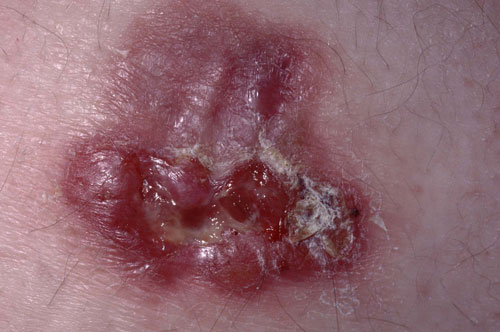
Anaplastic large cell lymphomas are a rare form of non-Hodgkin’s lymphoma which make up only 3% of lymphomas in adults and 10% in children. There are two different forms. The primary cutaneous type only affects the skin but the systemic type also affects the lymph nodes and internal organs as well. The condition occurs in people of all ages, but is seen most frequently in those aged 45 to 60, and is more common in men than women. There are no known risk factors. Lesions are typically red nodules that may ulcerate and itch and can occur anywhere on the body. As the lesions grow very slowly the condition often takes time to diagnose. Diagnosis is made by skin biopsy and the findings may need to be interpreted by a specialist histopathologist. Around 5-15% of those with primary cutaneous disease will progress to the systemic form. Treatment is with chemotherapy and sometimes radiotherapy. In the cutaneous type the prognosis is usually excellent.
Amelanotic melanoma
Malignant melanomas are typically irregularly pigmented lesions with large amounts of melanin present. Early diagnosis and excision is the mainstay of management. In the much rarer amelanotic melanoma the absence of melanin means that the lesions are much more difficult to diagnose. Appearing as pink or pale nodules, which may ulcerate, they are often mistaken for squamous cell carcinomas or pyogenic granulomas. The lack of pigment also means that many of the dermoscopic features of a pigmented melanoma are usually absent. This may result in an inappropriate biopsy technique being used that may delay diagnosis. Once diagnosed the treatment and prognosis are the same as for any other melanoma.
Dermatofibrosarcoma protuberans
This is a very rare malignant type of fibrosarcoma which develops in the dermis and affects one in a million people per year. In many respects it behaves like a benign lesion but the 2-5% risk of it metastasising means that it is managed as a malignant tumour. Dermatofibrosarcoma protuberans accounts for 2-6% of all soft tissue tumours and although most common in adults in their 30s
it can occur in children. The lesions typically present as a firm area of skin 1-5 cm in diameter, usually on the torso, which grows slowly. Diagnosis is made by an incisional biopsy of the lesion. Treatment is initially by excision, ideally by Mohs micrographic surgery, followed by chemotherapy or radiotherapy for the more aggressive tumours.
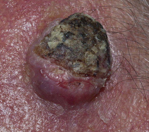
Squamous cell carcinoma
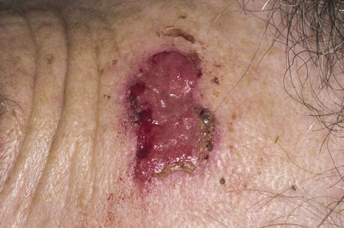
Squamous cell carcinomas (SCC) are the second most common type of skin malignancy after basal cell carcinomas (BCC) and classically present as a shallow ulcer with heaped-up edges often covered with a plaque. Unlike BCC which do not metastasise, SCC can spread to regional lymph nodes and may be fatal if left untreated. The incidence is on the increase worldwide. Increasing longevity and the use of sun beds, and environmental factors such as changes in the ozone layer are thought to be contributory factors. Patients on immunosuppressive medication following organ transplantation are also at increased risk and are usually monitored via a surveillance programme. If SCC is suspected referral should be made to secondary care via the two week wait pathway. Confirmation requires a biopsy after which wide excision is usually undertaken with skin grafting if appropriate. In those cases in which surgery is not possible radiotherapy can be undertaken.
 Download this article in PDF format