Haematoma
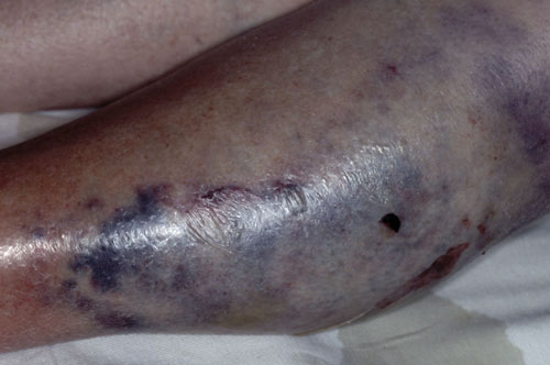
A haematoma is a localised collection of blood outside the blood vessels. Haematomas can develop in skin or other tissue such as muscles and may present as a hard mass. They vary in size from small bruises to large haematomas such as the one illustrated. With time the extravasated blood will be resorbed, initially this will be the serous fluid leaving the mass of blood cells and haemoglobin which may take many weeks to disappear. When the haematoma is in a muscle massage and exercise may help.
Furuncle
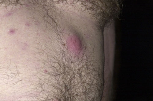
A furuncle is an infection, most commonly caused by Staphylococcus aureus, which develops deep in a hair follicle. The affected skin will feel hot, tender and swollen with erythema and a localised collection of pus. In more severe cases this may be associated with a fever and local lymphadenopathy. Furuncles can vary in size from a few millimetres to several centimetres in diameter. When multiple furuncles develop in close proximity the resulting lesion is termed a carbuncle. Where there are recurrent furuncles other associated conditions such as diabetes, obesity, malnutrition, and haematological disorders or the use of immunosuppressive drugs may be contributory factors. Complications include localised scarring and septicaemia. Very rarely, the infection may spread to other areas resulting in osteomyelitis, endocarditis and pneumonia.
Prayer nodule
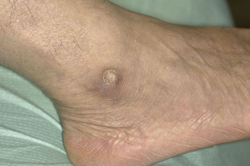
Prayer nodules occur as a result of repetitive trauma to localised areas of skin during regular prayers. The most commonly affected areas are the forehead, knees, ankles and the dorsum of the feet. One study looking at Muslims who prayed five times a day showed that these were present in 75% of men and 25% of women.1 The nodules are typically painless, well circumscribed, hyperkeratotic and hyperpigmented. Complications are rare but include secondary infection, ulceration and bleeding. Treatment includes surgical excision usually for cosmetic reasons or the application of 40% urea in white soft paraffin under occlusion followed by curettage. Recurrence can be reduced by trying to prevent further trauma to affected areas.
Reference
1 Abanmi AA, Al Zouman AY, Al Hussaini H et al. Prayer marks. Int J Dermatol 2002;41:411–4
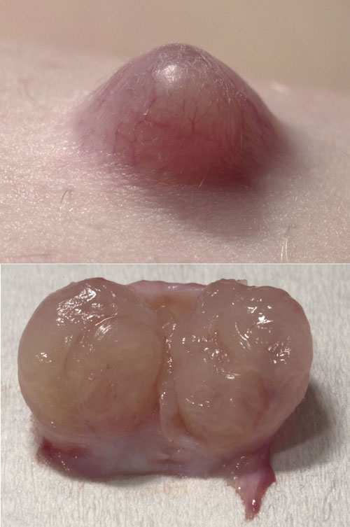
Pilomatrixoma
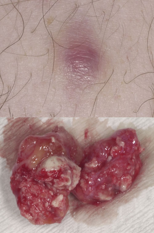
A pilomatrixoma, or calcifying epithelioma of Malherbe, is an uncommon benign hair follicle tumour derived from hair matrix cells. The presence of calcium within the nodule means that the lesions are characteristically bony hard. They often have an angulated shape the so-called tent sign. These lesions tend to occur on the head and neck but can develop in any area and are more common in young children. The cause is unknown. However, in affected hair cells an overactive proto-oncogene BCL-2 suggests that the normal process of cell death is suppressed and mutations in CTNNB1 imply loss of regulation of the beta-catenin/LEF protein complex. A pilomatrixoma may grow to several centimetres in diameter and the treatment of choice is surgical excision. Recurrence is common if the lesions are not completely excised.
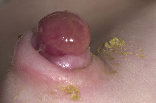
Umbilical granuloma
Umbilical granulomas develop at the base of the umbilicus after the cord has separated in the postnatal period. They are typically 1 mm to 1 cm in diameter and have a shiny, friable surface which may bleed on contact. They are usually light pink in colour. Darker red lesions may be umbilical polyps caused by retained gastric mucosa from the vitelline duct. Treatment is best undertaken by applying silver nitrate to the friable area. Usually more than one application is required to shrink the granuloma completely. Care is required to avoid contact with normal skin as silver nitrate may cause a burn. Those lesions which do not respond to silver nitrate may have an underlying umbilical sinus which requires surgical exploration and excision.
Neurofibroma
A neurofibroma is a benign peripheral nerve sheath tumour most commonly found in people with neurofibromatosis type 1. This is an autosomal dominant condition which occurs in around 1 in 3,000 births. It is also associated with café au lait spots, hypertension, learning difficulties and behavioural problems. Neurofibromas are usually first seen in the teenage years around the onset of puberty with numbers increasing through life, they are occasionally tender, may sting and itch but are asymptomatic in the majority of cases. There is no risk of malignant transformation. Treatment is not usually necessary but if required includes excision, laser ablation and hyfrecation.
 Download this article in PDF format