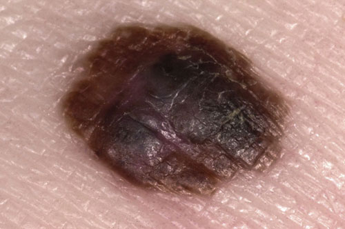
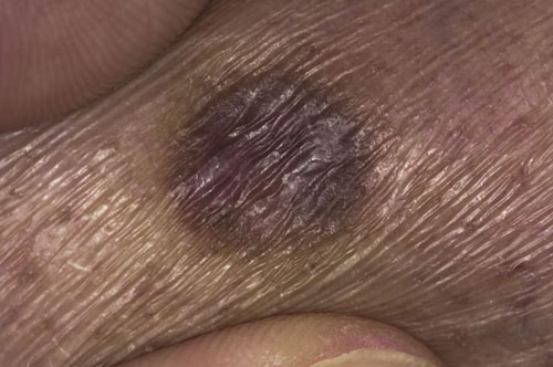
Malignant melanoma
Malignant melanomas are tumours of melanocytes and are associated with exposure to UV light. They are more common in individuals with Fitzpatrick skin types l and ll and in people who work outdoors. The incidence of melanoma has risen dramatically over the past few decades. In 10% of cases there is a positive family history, these patients typically develop melanomas at a younger age against a background of multiple dysplastic naevi. Diagnosis can be difficult and the use of a dermatoscope can help. Currently early surgical excision is the best form of treatment and to facilitate this suspected melanomas should be referred urgently under the two-week wait referral scheme to secondary care.
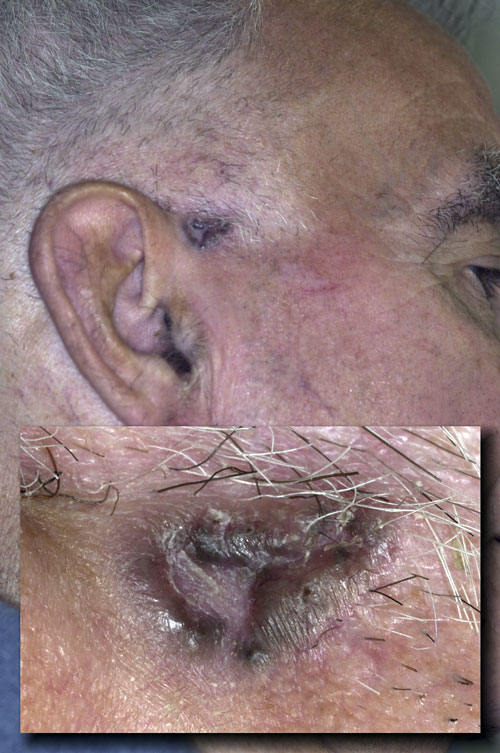
Pigmented basal cell carcinoma
Basal cell carcinomas (BCC) are the most common type of skin malignancy worldwide. Of these the nodular form, or rodent ulcer, is seen most frequently. In certain skin types these can be pigmented and in the pre-ulceration stage may be mistaken for melanomas. Pigmented BCCs are uncommon in white skin but in the Chinese population 80% of BCCs will be pigmented. The pigment varies from dark brown to black and tends to be seen in the thickest part of the BCC which is usually the rolled edges. Unlike melanomas where the whole lesion should be removed, a biopsy can be undertaken to confirm the diagnosis of a BCC prior to full excision. This can be helpful with large lesions where the diagnosis is in doubt. Pigmented BCCs are treated in exactly the same way as non-pigmented nodular BCCs with excision or radiotherapy.
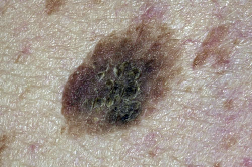
Seborrhoeic keratosis
Seborrhoeic keratoses are benign skin lesions which are increasingly common with age. They can be any colour from white through to black and darker lesions are often mistaken for malignant melanomas. They occur most frequently on the upper body especially the back and typically have a stuck-on, wart-like appearance, as only the top layer of the epidermis is involved. There are usually multiple lesions that vary in size from very small to 2-3 cm across. They may bleed when traumatised and feel rough to the touch. Treatment is not usually necessary but if required cryotherapy shave and cautery or shave excision can be helpful. As there is no viral component, wart treatments such as topical salicylic acid are of no help.
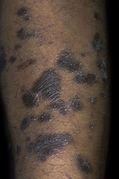
Lichen planus
Lichen planus is an unusual condition which can affect the skin, nails, scalp, genitals, and in 50% of cases the buccal mucosa. On the skin it forms very pruritic papules often described as violaceous. The size of the lesions varies from a few millimetres to over a centimetre. In individuals with Fitzpatrick skin types V or VI the lesions may be darker than the surrounding skin. A rare variant called lichen planus pigmentosus inversus presents with macules mainly in the axillae and never affects the scalp, nails or mouth. A skin biopsy can be useful to confirm the diagnosis if in doubt. Treatment is not always necessary but potent topical steroids can be helpful to decrease the pruritis. In more severe cases, other agents such as acitretin, methotrexate, hydroxychloroquine and phototherapy can be helpful. In 85% of cases the condition will have cleared after 18 months.
Dermatofibroma
Dermatofibromas are very common benign skin tumours usually found on the limbs. They are four times more common in females than males and can occur at any age. The aetiology is unclear but they appear to develop after localised skin trauma such as an insect bite, or inflammation from an ingrowing hair which causes clonal proliferation of dermal dendritic histiocyte cells. It is quite common for the nodule to develop pigment both centrally and in a perilesional distribution which may cause concern. Overlying skin is usually smooth but is tethered to underlying structures giving rise to the dimple sign whereby the surface dimples when lateral pressure is applied. If required they can be excised but treatment is not usually necessary.
Post-inflammatory hyperpigmentation
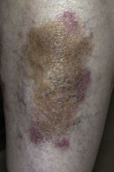
Any inflammatory skin condition involving the dermo-epidermal junction can cause hyperpigmentation. Conditions such as eczema, psoriasis, lichen planus and dermatitis are common causes. The pigment is usually more noticeable in those with Fitzpatrick skin types lV to Vl. Post-inflammatory hyperpigmentation typically has a dirty brown appearance unlike haemosiderin pigmentation associated with purpura and chronic venous insufficiency which is usually darker. In this case, the increased pigment was the result of underlying varicose eczema. Management should focus on the treatment of the underlying condition along with UV protection with sunscreens that will prevent a darkening of the pigment in the summer months.
 Download this article in PDF format