Impetigo
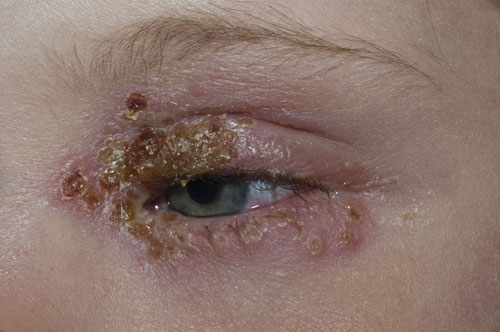
Impetigo is a highly contagious bacterial skin infection caused by Staphylococcus aureus or Streptococcus pyogenes. It is most common in young children and often occurs in outbreaks. Impetigo is spread
by direct contact with an infected individual. It can affect any area of the body including the skin of the eyelids. Differential diagnoses include herpes zoster, eczema, erysipelas, and necrotising fasciitis. Treatment is with either topical or oral antibiotics. In this case the close proximity to the eye meant that an oral antibiotic flucloxacillin was the treatment of choice.
Molluscum contagiosum
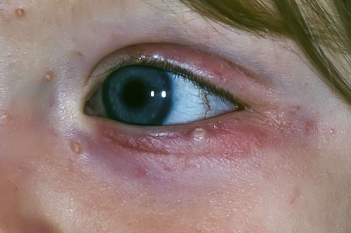
Mollusca are caused by infection with a virus from the pox family. It is a very common infection in childhood. Lesions are typically dome shaped with a central depression varying in size from 1 mm to 1 cm in diameter. Lesions can occur anywhere on the body including the peri-ocular skin where diagnosis can be more difficult especially if there is only a single lesion. In this case, the presence of multiple umbilicated lesions around the eye was a clue to diagnosis. Treatment is not usually necessary as spontaneous remission tends to occur. In severe cases liquid nitrogen or curettage and cautery may be used. Care should be taken when the eyelids are involved especially as scarring may adversely affect lid function.
Pterygium
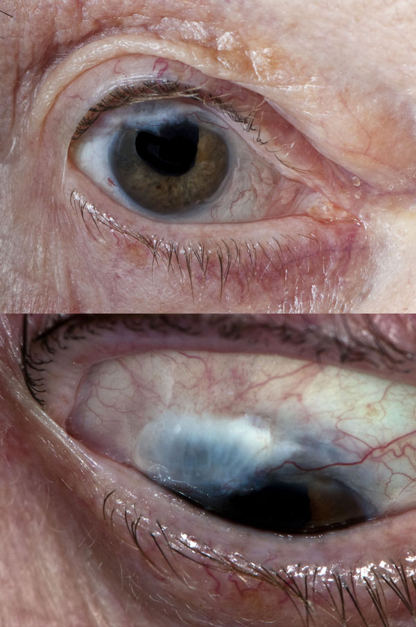
A pterygium is a benign lesion on the conjunctiva, most commonly growing from the nasal side of the sclera. It is thought to be associated with exposure to ultraviolet light, low humidity, wind and dust, hence it is sometimes referred to as surfer’s eye. It is characterised by elastotic degeneration of collagen and fibrovascular proliferation. Wearing sunglasses and using artificial tears may prevent the lesion from developing further. In more severe cases the eye may feel inflamed and sore. If the pterygium grows across the pupil vision may be affected and surgery required. As there is no reliable medical treatment this is usually the only option. However, following surgery the condition may recur, hence some cases require repeated surgery.
Subconjunctival haemorrhage
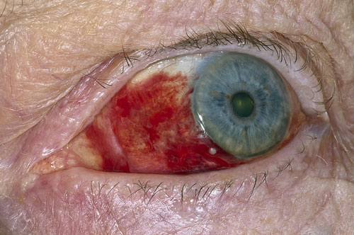
Subconjunctival haemorrhage results from bleeding of the conjunctival or episcleral vessels into the subconjunctival space. This may occur spontaneously, following trauma, or secondary to hypertension or disorders that affect clotting. Although it can occur at any age subconjunctival haemorrhage is more common with increasing age and may be a recurrent condition. It is also more common in patients with diabetes, hyperlipidaemia or ischaemic heart disease. It is usually asymptomatic or associated with mild irritation. As with any bruise the bright red appearance will fade to a green or yellow colour before resolving completely. Be vigilant for a haemorrhage which does not have a visible posterior margin as it may be a sign of an intracranial bleed or orbital roof fracture.
Corneal rust spot
After entering the eye particles of steel or iron will often rust leaving either a ring or a rusty mark. In this case the patient had been using a grinder. He inspected the eye himself but no foreign body was visible. However, three days later the rust made the foreign body more obvious. If in doubt fluorescein drops can help locate a foreign body or a laceration through which a foreign body may have passed into the eye itself. In this case the metal was removed at the eye casualty department under local anaethesthetic, and antibiotics drops were prescribed. Failure to do this can risk corneal scarring which may affect the patient’s eyesight.
Basal cell carcinoma
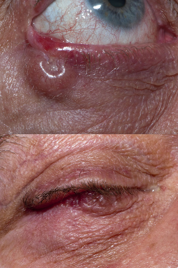
Basal cell carcinoma is the most common type of skin cancer in the UK. Lesions occur predominantly on areas exposed to ultraviolet light and do not metastasise. Most lesions are easily treated by surgical excision. If a basal cell carcinoma develops on the eyelids treatment can be more difficult and referral to an oculoplastic surgeon is required. In this patient the basal cell carcinoma was on the lower lid and was treated with a Hughes flap, seen in the image below. In this instance the edge of the lesion is easily discernible. However, in other cases it may not be as clear. In such cases Mohs surgery is the treatment of choice where the tissue is examined immediately after removal to check for clearance. Cure rates of over 95% have been reported with this technique even in recurrent tumours.
 Download this article in PDF format