Skin metastases
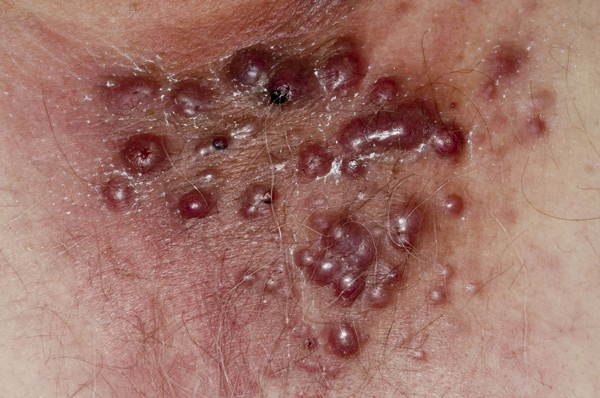
Internal malignancies metastasise to the skin in 3-10% of cases. Although this usually occurs late in the disease process and is associated with a poor prognosis, it will sometimes be the first indication of an underlying malignancy. In such cases a biopsy of the skin deposit may help identify the location of the primary tumour.
The most common malignancy to spread to the skin is breast cancer, although in this case the primary was in the lung. Other tumours that metastasise to the skin include those of the GI tract, uterus, prostate and kidney with the most frequently affected areas being the umbilicus and scalp. When scalp metastases develop the first sign is usually localised alopecia which will be scarring often with telangiectasia present.
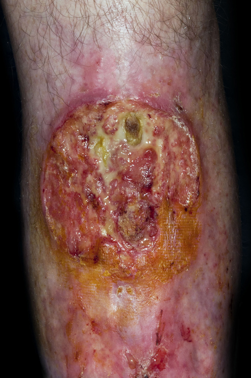
Pyoderma gangrenosum
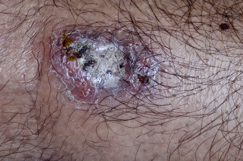
Pyoderma gangrenosum presents with an inflamed nodule or pustule which ulcerates centrally and then expands typically with a polycyclic or serpiginous outline and a blue edge. Lesions may be single or multiple and can occur anywhere on the body. The pathogenesis of the condition is not fully understood, an immunological process is thought to be involved. It is commonly associated with an underlying condition such as inflammatory bowel disease, rheumatoid arthritis, leukaemia and monoclonal gammopathies. Often treating the underlying condition e.g. Crohn’s disease will improve the lesions of pyoderma, but in general they will respond to oral steroids and will heal leaving papery scars.
Xanthelasma
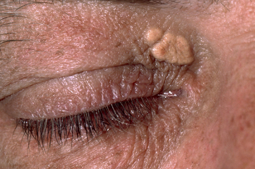
Xanthoma are deposits of fatty material in the skin and subcutaneous tissue. One of the most common places for these to form is the eyelids where they are referred to as xanthelasma palpebrarum. The lesions are sometimes associated with disorders of lipid metabolism, and patients should have their lipid levels checked. They are most commonly seen in types ll, lll and lV hyperlipidaemia. Treating raised lipid levels with statins will reduce the risk of developing further xanthoma, but will not reduce lesions already present. If required, these can be treated with topical trichloroacetic acid, direct destruction with a hyfrecator, cryotherapy, or carbon dioxide or argon laser therapy.
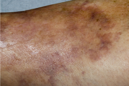
Erythema nodosum
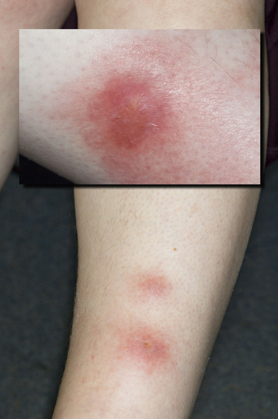
Erythema nodosum results from an immunological reaction to various triggers causing inflammation of subcutaneous fat.
Triggers include infection (bacterial, viral or fungal), malignancy, drugs, or systemic diseases such as sarcoidosis, ulcerative colitis and Crohn’s disease. Erythema nodosum presents with tender nodules on the legs, forearms or breasts, which are bruise like in appearance, and may be associated with fever and generalised arthralgia.
The lesions typically last for six to eight weeks with new lesions appearing and resolving over this period. Management involves a careful history and examination followed by investigations to find the cause. Once the cause is dealt with the condition will improve. In the interim NSAIDs may be helpful.
Graft versus host disease
Acute graft versus host disease (seen here) will occur within four weeks of bone marrow grafting for conditions such as leukaemia and aplastic anaemia. It is caused by donor lymphocytes reacting against host tissues in areas such as the skin, bowel and liver. Patients report fever and generalised malaise along with a progressive rash which may progress to epidermal necrolysis. Chronic graft versus host disease takes longer to develop and causes a skin rash which looks like scleroderma or lichen planus. Treatments include oral steroids, azathioprine and ciclosporin A.
Necrobiosis lipoidica
Although less than 1% of diabetes patients will develop necrobiosis lipoidica, most people with the condition will also have diabetes or impaired glucose tolerance. Necrobiosis lipoidica occurs on the shins with either single or multiple plaques. The lesions are typically shiny and atrophic, reddy brown-yellow in colour with underlying blood vessels visible through the skin. Affected areas become very vulnerable to ulceration which may occur after minor trauma to the area. Healing of ulcers can be very slow. The condition tends to be chronic with no reliable treatment available.
 Download this article in PDF format