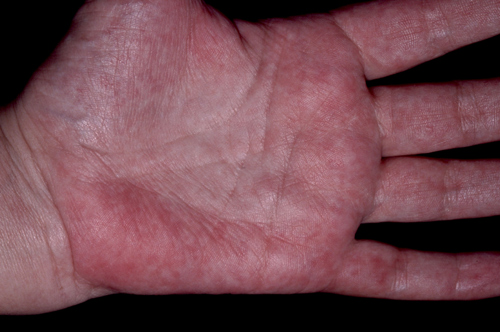
Palmar erythema
Palmar erythema is a reddening of the palms, especially the thenar and hypothenar eminences. It can exist as a primary physiological condition but may also be secondary to an underlying disease. It is seen in about 30% of pregnant women and 23% of patients suffering from cirrhosis of the liver. Other causes include chronic liver disease, connective tissue disorders, thyrotoxicosis and leukaemia. Drug-induced palmar erythema has been reported, with hepatic damage, with drugs such as amiodarone, gemfibrozil and cholestyramine; and without hepatic damage in the case of topiramate and salbutamol.
Varicose veins
The primary cause of varicosities is incompetent valves within the veins. Traditionally treatment has consisted of removal or obliteration of the great saphenous vein by ligation and stripping techniques. In recent years the development of endovascular lasers has revolutionised management enabling the varicosities to be treated from within the vessel. The laser is placed in the lumen of the vessel and passes along its length. It is positioned using ultrasound or a guide light. A tumescent solution with local anaesthesia is injected around the entire length of the vessel which protects adjacent structures such as nerves or other vessels from the laser. It also helps with pain control. External pressure is applied to collapse the vein around the laser which is then fired producing heat, intraluminal steam bubbles and thrombosis, which in turn destroys the endothelium. The laser is slowly withdrawn and the procedure repeated until the entire length of the vein has been treated.
Vasculitis
Vasculitis is a reactive inflammatory condition of the blood vessels which may affect both the skin and internal organs. The rash is typically non blanching. Clinical presentation will vary depending on the histological type of inflammation, the vessels involved and their distribution. Vasculitis can be classified according to the size of the vessel affected: large vessel disease e.g. polymyalgia rheumatica, medium vessel e.g. polyarteritis nodosum or small vessel e.g. Henoch Schonlein purpura. Other classification systems are based on the cause or the location of the affected vessels. In this case, the cause was an allergic vasculitis in which the superficial dermal capillaries, venules and arterioles are affected. Fibrinoid necrosis of vessels and surrounding tissues has occurred with extravasation of red blood cells. Associated renal involvement should always be considered and urine checked for the presence of blood. Complete recovery usually occurs, although the prognosis depends on the extent of any renal involvement associated with the rash.
Erythema ab igne
Erythema ab igne is caused by chronic exposure to moderate heat from an external source such as a fire or hot water bottle. The reticulate erythematous patches may be associated with telangiectasia and hyperpigmentation. It most commonly affects the legs of women in the 40 to 70 age group and patients with chronic pain such as pancreatitis and low back pain. The differential diagnosis includes livedoid vasculitis and poikiloderma atrophicans vasculare. Once the heat source is removed most cases will resolve over a few months. For those that do not, treatment options include photothermolysis using the Nd:YAG, ruby, or alexandrite lasers or alternatively camouflage make up may be applied. In chronic cases malignant transformation into squamous cell carcinoma may occur but this is very rare.
Spider naevi
These stellate telangiectases look like spiders with multiple vessels branching from a single central feeding vessel. Pressure to these vessels will result in the disappearance of the whole lesion which will reappear when the pressure is relaxed. They are common on the faces of children and in women may erupt during pregnancy. If multiple spiders erupt in the distribution of the superior vena cava this may be a presenting sign of liver disease and further investigations including liver function tests should be undertaken. Spider naevi do not require treatment except for cosmetic reasons when a fine-tipped hyfrecator may be applied to the central vessel, usually without the need for anaesthesia.
Angiokeratoma of Fordyce
First described in 1896 by John Fordyce, scrotal angiokeratomas are usually multiple and asymptomatic. Their size can vary between 2 and 5 mm and they may extend onto the penis and adjacent areas of the thighs or abdomen. Scrotal angiokeratomas are composed of ectatic thin-walled vessels in the superficial dermis with overlying epidermal hyperplasia. The differential diagnosis includes cherry haemangioma, genital warts, granuloma pyogenicum, squamous cell carcinoma and Fabry syndrome. Treatment is not usually required unless the lesions bleed, or for cosmetic reasons. In these cases cautery with a hyfrecator is usually well tolerated. Alternative approaches include cryotherapy, excision and laser treatment.
 Download this article in PDF format