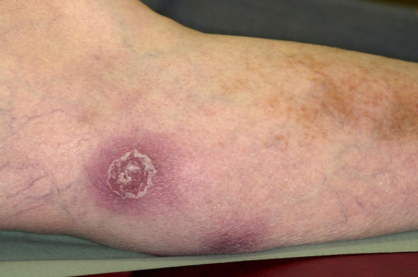
Discoid lupus erythematosus
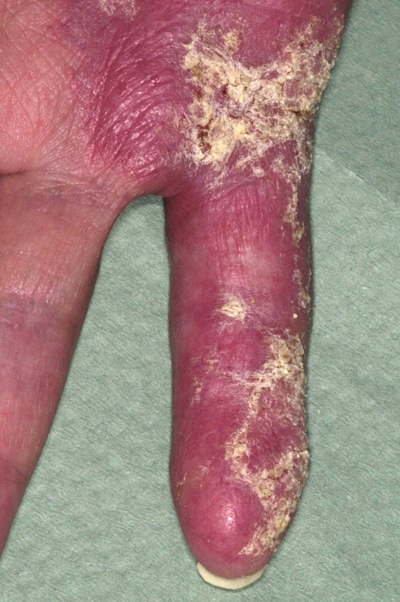
Discoid lupus erythematosus (DLE) is a rare chronic condition confined to the skin. It forms part of the wide range of lupus erythematosus conditions some of which affect internal organs. The cause is not fully understood but the rash gets worse when affected skin is exposed to sunlight. Affected areas may be itchy or tender especially on fingers but generally are asymptomatic. The diagnosis is confirmed by skin biopsy. Treatments include sunscreens, strong topical steroids and hydroxychloroquine which bring the condition under control but are not curative. Around 5% of people with DLE will develop systemic lupus erythematosus (SLE).
Dermatomyositis
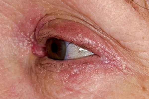
Dermatomyositis is an idiopathic inflammatory condition which most commonly affects skin and muscles but may also affect the joints, oesophagus, lungs and heart. There are various cutaneous manifestations including the heliotrope facial rash, Gottron’s papules, poikiloderma and scalp lesions. The heliotrope rash is highly suggestive of the condition and consists of a violaceous or dusky erythematous rash affecting the periorbital skin with or without oedema. In some cases it may be a subtle sign with only a mild discoloration along the eyelid margins. A skin biopsy can aid diagnosis but histological changes may be difficult to differentiate from the changes seen in lupus. It is important for patients to avoid sunlight and sunscreens should be used. Corticosteroids may also be helpful.
Scleroderma
Scleroderma is a systemic disease characterised by skin thickening and induration which is the most obvious feature. Internally the condition causes tissue fibrosis and chronic inflammatory infiltration of the GI tract, lungs, kidneys, cardiovascular and genitourinary system.
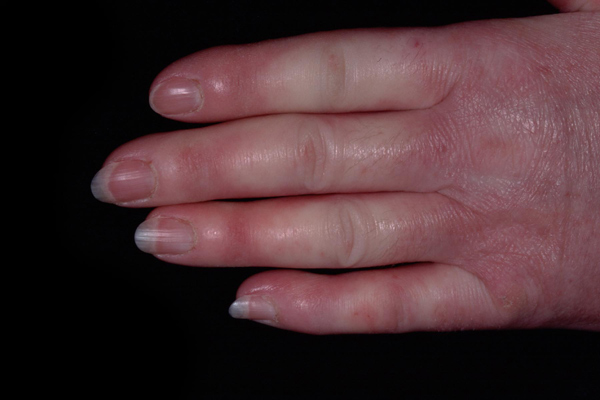
In some cases it can be fatal, often as a result of pulmonary hypertension and pulmonary fibrosis. It is more common in women and has a peak age of onset between 50 and 60. Skin signs include skin tightness leading to a beak-like appearance, sclerodactyly with digital ulceration, loss of skin creases, joint contractures and sparse hair. Salt and pepper hypopigmentation and diffuse pruritus also occur. Around 70% of patients initially present with Raynaud’s phenomenon with 90% going on to develop it during the course of their disease.
Morphoea
In morphoea, also known as localised scleroderma, excessive collagen is deposited in the dermis or subcutaneous tissue giving the skin a thickened appearance. There are four subtypes (circumscribed, generalised, linear and pansclerotic) which differ in presentation and depth of tissue involvement. Although morphoea is not associated with the systemic features seen with scleroderma, it can be associated with fever, lymphadenopathy, arthralgia, and fatigue. Between 0.9 and 5.7% of patients will progress to scleroderma. A biopsy can be helpful in the diagnosis but changes are similar to those seen with scleroderma. Lesions generally undergo gradual spontaneous regression over a 3-5 year period. With localised disease, topical superpotent steroids, imiquimod and tacrolimus can be helpful in active lesions. In more disabling disease, aggressive therapy with oral steroids, mycophenolate, hydroxychloroquine or phototherapy may be required.
Lichen sclerosus
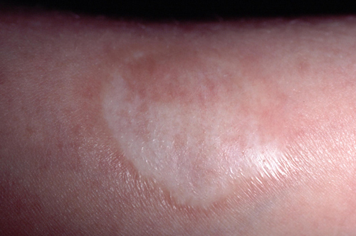
Lichen sclerosus is a chronic inflammatory dermatosis in which epidermal atrophy and scarring produce white plaques. Although more common on the genitals, extragenital manifestations can also occur, most often on the back and shoulders. Affected areas are usually asymptomatic but may be itchy. The male to female ratio is 1:6. and up to 15% of cases occur in children. The aetiology is unknown but may include genetic, infectious, hormonal and environmental factors. Where the diagnosis is in doubt a small biopsy may be helpful, and should be taken from the most longstanding area affected. Treatment is not usually required for extragenital lesions though antipruritics can be helpful in symptomatic cases.
Panniculitis
Panniculitis describes a group of conditions which involve inflammation of subcutaneous fat. Clinically they all have a similar appearance, but histologically they have characteristic features which can be useful in differentiating between the different types. Affected skin typically feels thickened when touched with a reddened or darker appearance and is usually tender, forming raised nodules. Treatment approaches include rest and elevation of affected areas, compression hosiery, analgesia and systemic steroids. Severe persistent localised areas may require surgical excision.
 Download this article in PDF format