Cylindroma
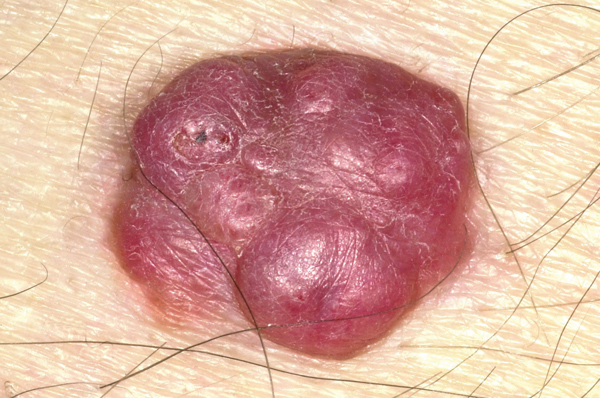
A cylindroma is a rare, benign adnexal tumour which is thought to arise from the apocrine or secretory coiled duct. In this case, a solitary lesion was present. However, in the case of Brooke-Spiegler syndrome or turban tumour syndrome multiple cylindromas are seen in close proximity and may coalesce. They occur most commonly on the head and neck from middle age onwards. The differential diagnosis includes: pilar cysts, eccrine spiradenomas and follicular infundibulum tumours. Surgical excision is the treatment of choice for solitary lesions.
Cutaneous B-cell lymphoma
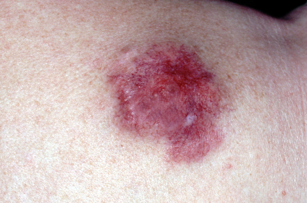
B-cell lymphomas are the most common of the nodal lymphomas accounting for 20-25% of all cutaneous lymphomas. Overall these lymphomas have a good prognosis if recognised early. The aetiology is not clearly understood and is thought to be a multifactorial and multistep process. The differential diagnosis includes T-cell lymphoma and B-cell pseudolymphoma. Diagnosis is confirmed by histological examination of a skin sample obtained by punch biopsy. Treatments include surgical excision, antibiotics (doxycycline 1 mg twice a day for three weeks), and radiotherapy. Second-line treatments include intralesional steroids, rituximab or interferon alpha.
Cutaneous lava migrans
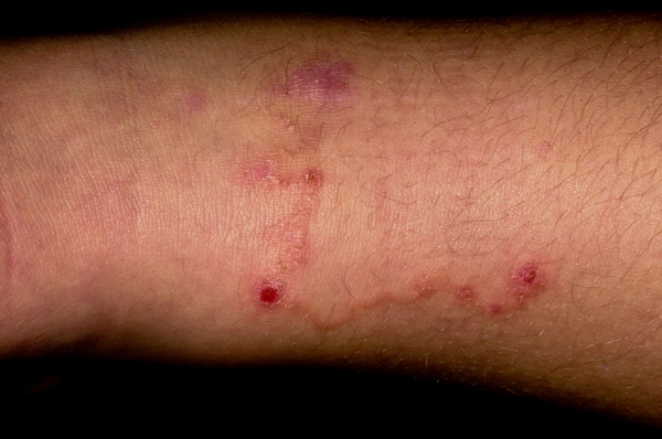
Cutaneous lava migrans is the most commonly acquired tropical dermatosis. It has a very characteristic serpiginous rash which is typically pruritic, erythematous and slowly advances if not treated. It is the result of a parasitic infestation which begins when eggs from animal faeces are deposited usually on sandy soil or a beach. These eggs hatch and the larvae feed on bacteria until they come into contact with skin which they penetrate through follicles, fissures or directly through intact skin. Once in the skin of a suitable host, they penetrate through the dermis into the lymphatics. They then migrate to the lungs where they break through the alveoli and move into the trachea from where they are swallowed and excreted in faeces. The condition is usually self-limiting and treatment is not necessary. If required, oral thiabendazole is the treatment of choice. Cryotherapy can also be useful but needs to be targeted a few millimetres ahead of the rash which is where the parasite is usually positioned.
Lupus pernio
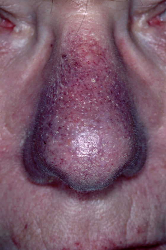
Lupus pernio is a chronic granulomatous condition characterised by raised indurated lesions of the skin which are usually purple in colour. The most commonly affected areas are the nose, ears, cheeks, lips, fingers and forehead. It is the most characteristic skin manifestation of respiratory sarcoidosis. Although it is not common in patients with sarcoidosis it is pathognomonic of the disease.
In recent years, trials using adalimumab and infliximab have proved promising in the treatment of this condition which has relied on systemic steroids.
Yellow nail syndrome
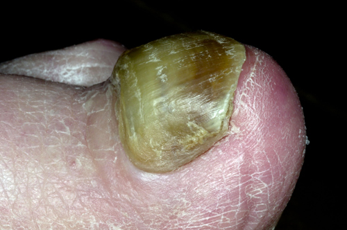
Yellow nail syndrome is a rare condition which is usually accompanied by lymphoedema. In some cases it is also associated with recurrent pleural effusions and less commonly bronchiectasis, chronic bronchitis and sinus infections. It generally occurs after middle age, but is occasionally seen in children. All the nails are usually affected, becoming thickened and static with little or no new growth. Cuticles tend to be lost and onycholysis may occur. Management includes treatment of any underlying conditions, but there is often little that can be done to help the nails and the changes are usually permanent. In rare cases, treatment of the underlying condition will help reverse the changes seen in the nails.
Urticarial vasculitis
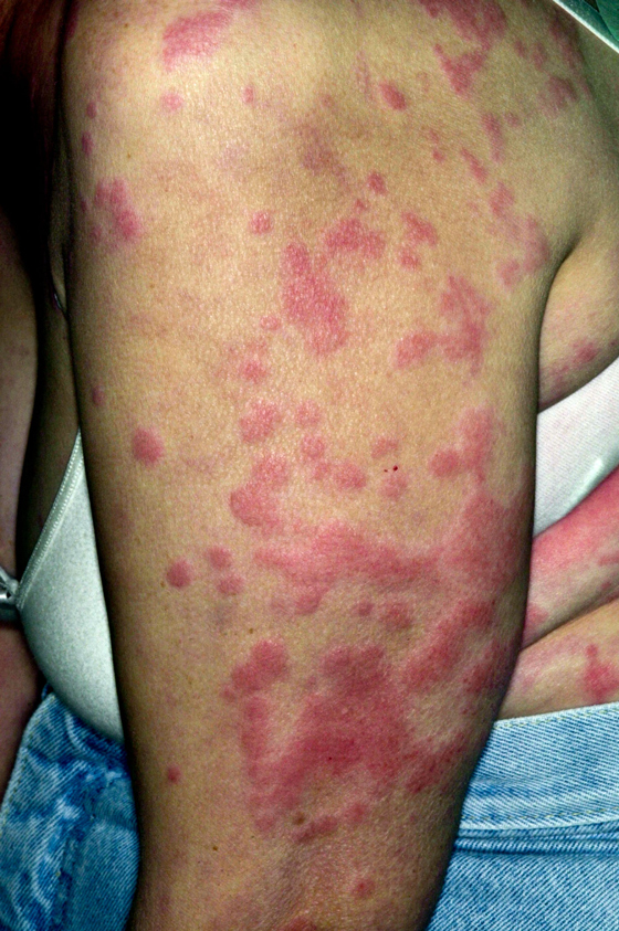
Urticarial vasculitis is similar in appearance to urticaria, but histologically has an underlying vasculitic component. The cause is unknown but it is associated with other conditions such as inflammatory connective tissue disorders e.g. SLE and Sjogren’s syndrome; leukaemia and internal malignancies; and viral disease such as hepatitis B and C. It can also be caused by certain drugs such as ACE inhibitors. The rash usually starts with a painful or burning sensation with pruritus followed by typical urticarial red patches. Unlike urticaria these last for more than 24 hours, slowly resolving and leaving behind areas of bruising or hyperpigmentation. The diagnosis can be confirmed by skin biopsy. Treatments include antihistamines, NSAIDs, dapsone, colchicine, hydroxychloroquine and oral steroids.
 Download this article in PDF format