Basal cell carcinoma
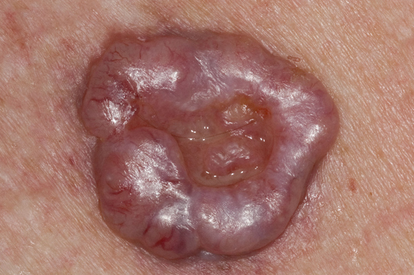
Nodular basal cell carcinoma (BCC) is the most common type of skin cancer. BCCs form initially as translucent skin-coloured papules which expand laterally into annular lesions with a typical pearly, raised edge and central ulceration. They are also known as rodent ulcers because of their resemblance to rat bites. Closer inspection will reveal fine telangiectatic blood vessels running over the surface of the tumour. BCCs are typically very slow growing expanding to 1-2 cm over five to ten years if left untreated. The treatment of choice is surgical excision. Other options include radiotherapy, cryotherapy and curettage and cautery. Incomplete excision increases the risk of recurrence which can be more difficult to treat. Mohs micrographic surgery, where available, is the treatment of choice for recurrences. It has a 95% cure rate at five years.
Erythema annulare centrifugum
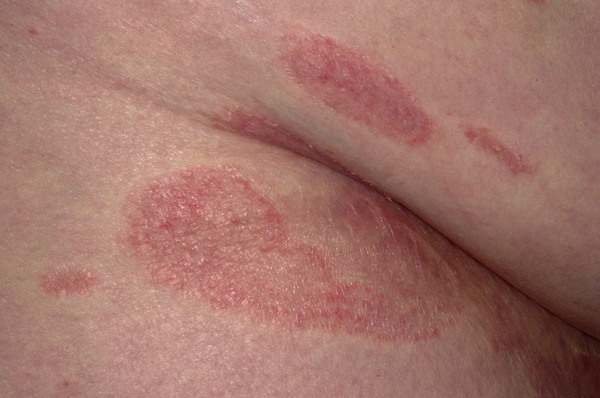
Erythema annulare centrifugum usually presents as an asymptomatic or pruritic annular rash which may be associated with an underlying condition such as an infection or malignancy. It may also be caused by certain drugs, for example, cimetidine, spironolactone, salicylates, penicillin and amitriptyline. The rash will normally start with a localised crop of papules which spread laterally at a rate of 2-5 mm per day producing a red-coloured annular rash which will then clear centrally. The thighs and legs are most commonly affected. The condition is usually self-limiting, especially if an underlying cause is found and either treated or removed. In symptomatic cases topical, oral or intralesional steroids can be effective.
Erythema multiforme
Erythema multiforme usually occurs as a reaction to a drug or an infection such as herpes simplex. Typically annular non-scaling plaques or target lesions will develop on the hands, feet, forearms and legs. Occasionally lesions will blister and may develop into the more severe variant of Stevens-Johnson syndrome. In general, new lesions will develop for one to two weeks until the cause has been removed or the underlying infection has resolved. Individual lesions last for one to two days. The condition is best managed by identifying and removing the cause. Where the cause is recurrent herpes simplex infection, such as cold sores, prophylactic oral aciclovir may be required daily. If a drug cause is suspected the drug should be withdrawn and avoided in future. Once the rash has formed symptomatic treatment with antihistamines can be helpful.
Localised granuloma annulare
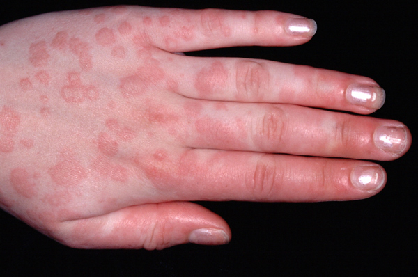
Granuloma annulare is a relatively common benign dermatosis. The condition can occur at any age, the cause is unknown. As it arises from the dermis, rather than the epidermis, affected areas have no scale or crusting. There are a number of different varieties including: localised; generalised; subcutaneous; perforating and arcuate. In localised granuloma annulare, the lesions are asymptomatic, most commonly affecting the dorsal surfaces of the hands, feet and fingers. Lesions first appear as small papules which join up to form annular plaques 1-5 cm in diameter. Treatment is not usually required as the lesions will resolve spontaneously. Painful or unsightly lesions can be treated with potent topical steroids or intralesional steroid injections.
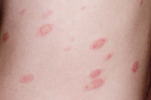
Pityriasis rosea
Pityriasis rosea starts with the development of an annular herald patch, 2-5 cm in diameter, usually on the trunk. A few days later this is followed by the appearance of multiple, small plaques which also occur mainly on the trunk in a Christmas tree distribution. The plaques all have a typical collarette of fine scale peripherally and may be slightly itchy. They usually resolve spontaneously over two to ten weeks leaving some postinflammatory hyperpigmentation. The condition is not contagious and is more common in the autumn and winter. No treatment is usually required, but a moderately potent topical steroid can be useful if the lesions are itchy. UVB phototherapy has also been shown to reduce itching and speed up the time to resolution.
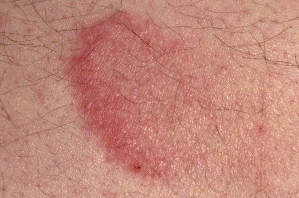
Tinea corporis
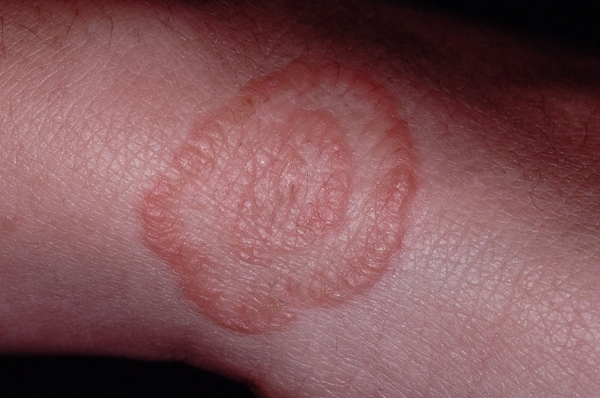
Tinea infections are usually caused by one of three types of fungus: Trichophyton; Microsporum or Epidermophyton. These invade the keratin of the skin where their metabolic products cause inflammation. With time tinea infections spread laterally causing annular lesions with a leading inflamed edge and central clearing, which gives rise to the term ringworm. Affected skin is typically very itchy, with scaling and erythema and, in some cases, pustules or vesicles. If the diagnosis is in doubt skin scrapings can be taken for microscopy and culture. In most cases topical antifungal creams such as clotrimazole will be adequate. In more severe cases an oral antifungal may be required.
 Download this article in PDF format