Vitiligo
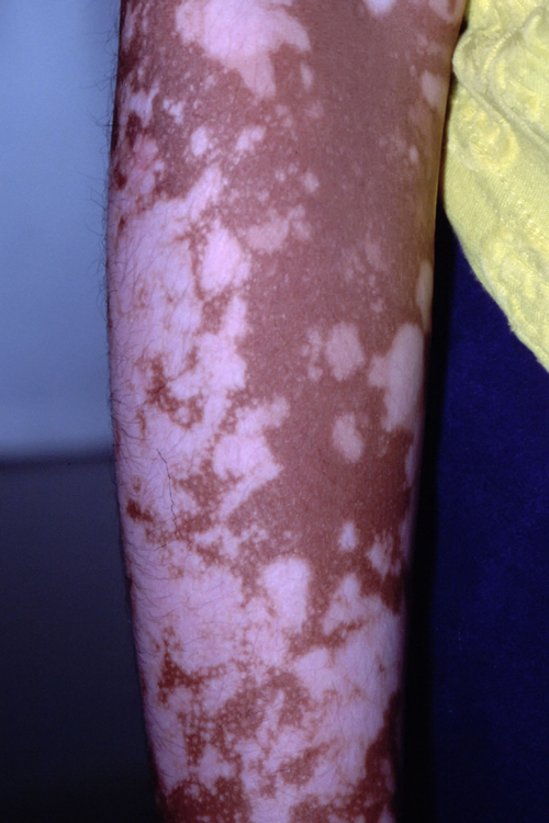
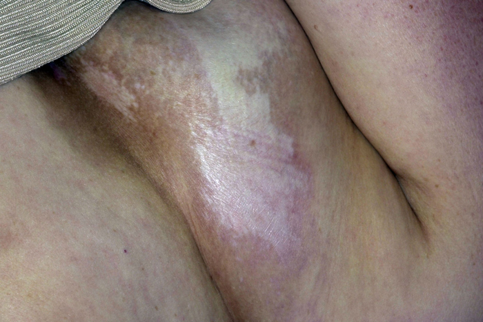
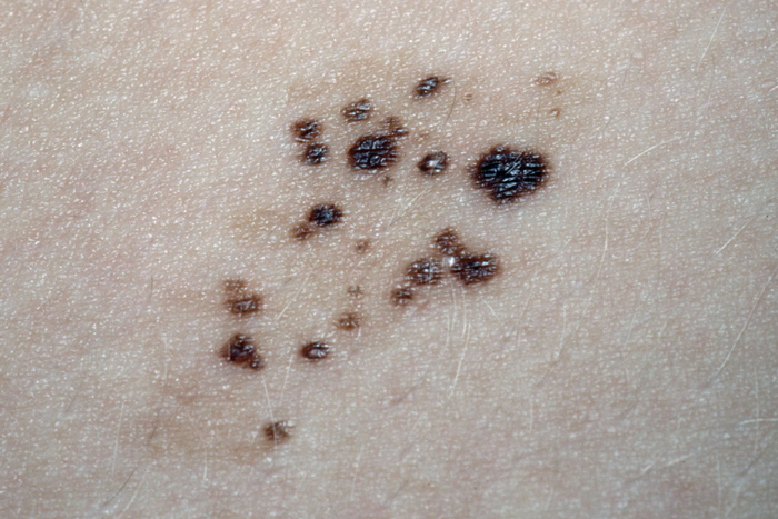
Vitiligo is an acquired condition in which progressive destruction of melanocytes results in loss of skin pigment. It affects 0.5-2% of the world population with an average age of onset of 20 years. The most commonly affected areas are the face, neck and scalp. Vitiligo tends to occur in areas of repeated trauma and scars. Treatment options include systemic phototherapy with psoralens, lasers and topical therapy with an immunomodulator such as tacrolimus. Management can be a challenge as none of these approaches works particularly well. Other treatments include depigmentation of remaining pigmented areas to give an even colour. As this is permanent, counselling is essential before treatment is undertaken. In all cases, the use of high factor sun screens will help reduce the contrast between affected and unaffected areas. The psychological impact of vitiligo must never be underestimated especially in darker-skinned individuals.
Post-inflammatory hyperpigmentation
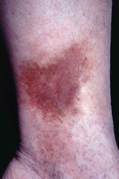
Areas of increased pigmentation can develop in normal skin for a variety of reasons. Inflammatory conditions, infections, eruptions, allergic reactions, trauma e.g. burns, and mechanical injuries can all result in hyperpigmentation. Post-inflammatory hyperpigmentation (PIH) can be particularly problematic for darker-skinned individuals with acne or eczema. Once the areas of pigment develop treatment can be difficult and the skin may be permanently affected. When considering a diagnosis of PIH a careful drug history should be taken as certain drugs e.g. amiodarone, amitriptyline and minocycline can cause pigment changes within the skin. As with many other disorders of pigmentation, treatment can be difficult and it may be many months before an improvement is noticed. Where an underlying condition, such as acne or eczema, has been identified as the cause it should be treated as soon as possible to prevent further hyperpigmentation. Topical treatments include tretinoin cream, glycolic acid, azelaic acid, corticosteroids, and hydroquinone.
Acanthosis nigricans
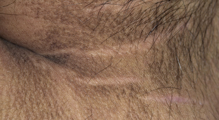
Acanthosis nigricans is an asymptomatic darkening and thickening of the skin. It starts as hyperpigmented macules, which may be pruritic, and then progresses to palpable plaques. The rash is symmetrical and has a velvety feel. The most commonly affected areas are the axillae, groin and posterior neck. Acanthosis nigricans takes several forms, from a simple benign condition to one associated with underlying malignancy. In the latter, the rash appears before any signs of the cancer in a third of cases. Apart from underlying malignancy, other associated conditions include obesity, acromegaly, dermatomyositis, lupus, scleroderma, hyperinsulinaemia to name but a few. There is no treatment of choice, the goal is usually to treat the underlying cause. Treatment to improve the cosmetic appearance of affected areas includes dermabrasion, surgical excision and laser therapy.
Pityriasis versicolor
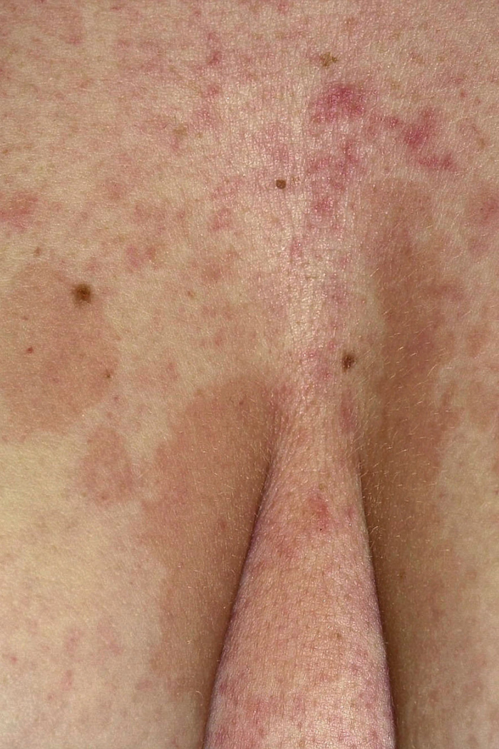
Pityriasis versicolor is a relatively common benign condition associated with the yeast Malassezia. Infection results in areas of either hypo- or hyperpigmentation usually over the trunk. For many individuals it only becomes apparent when they get tanned on holiday, and is often assumed to be the result of skin peeling. Closer examination reveals a fine dust-like scale over the affected areas. For most patients a topical antifungal shampoo applied as a lotion directly to the skin before showering will help. In more severe or resistant cases oral antifungal medication such as fluconazole or itraconazole may be required. It is important to explain that it may take many months for the affected areas to repigment. Absence of scale is a good indication that the treatment has been successful.
Lichen sclerosus et atrophicus
This chronic inflammatory dermatosis presents with white papules which coalesce into plaques with a shiny porcelain surface. Although lichen sclerosus et atrophicus is most commonly seen in the female genital area it can occur anywhere on the body and in either sex. Around 15% of cases occur in children, usually affecting the genitals. Extra-genital cases may be asymptomatic, but genital cases are usually intensely itchy. As the disease progresses the skin becomes sclerotic and tightens leading to obliteration of affected tissue on the labia or foreskin with a resultant loss of function. In chronically affected areas malignant change may occur. Affected extra-genital areas that are asymptomatic may require no treatment. However, for symptomatic genital disease potent topical steroids are usually required to slow disease progression. In males, circumcision may help. In females, surgery is not usually recommended unless malignant change is suspected.
Speckled lentiginous naevus
Speckled lentiginous naevus is a patch of darkly pigmented macules and papules that can occur anywhere on the body. It is thought to represent a localised defect in neural crest melanoblasts. Around 80% of these lesions are present at birth or appear in early infancy as pale café au lait macules with the darker papules appearing later. The size of the underlying macule can vary from a few centimetres to >60 cm in diameter and
may increase with time. Atypical transformation may occur in the darker papules leading to the development of cutaneous melanoma, so observation for change ideally with photography or mapping should be undertaken. Any suspicious areas should be excised for histological examination.
 Download this article in PDF format