SEARCH

Keteepe-Arachi T, Sharma S. Underestimating risk in women delays diagnosis of CVD. Practitioner 2016;260(1791):11-15
Underestimating risk in women delays diagnosis of CVD
21 Mar 2016
AUTHORS
Dr Tracey Keteepe-Arachi BSc (Hons) MBBS MRCP, Clinical Research Fellow and Cardiology Specialist Registrar
Professor Sanjay Sharma MD FRCP (UK) FESC (UK), Professor of Clinical Cardiology, St George’s, University of London, London, UK
Article
Abstract
CVD remains the most common cause of mortality in women. In 2007, the annual mortality in women secondary to CAD was 4.7 times that of breast cancer. Around 2.8 million women are living with CVD in the UK. There has been an increase in the prevalence of MI in women aged 35 to 54, while a decline in prevalence was observed in age-matched men.Difficulty in evaluating symptoms of ischaemic heart disease in women is well documented and remains challenging because of their atypical nature. The main gender difference is that women tend to present less frequently with exertional symptoms of chest pain before an AMI. Although men and women share classic cardiovascular risk factors the relative importance of each risk factor may be gender specific. The impact of smoking is greater in women than men, especially in those under 50. Diabetes is a more potent risk factor for fatal CHD in women than men. Risk factors specific to women include postmenopausal status, hysterectomy and complications during pregnancy. Women who develop gestational diabetes mellitus or pre-eclampsia more than double their risk of CVD later in life. Transition to the menopause is associated with a worsening CHD risk profile. After the menopause women may experience an increase in weight, alteration in fat distribution and an increase in other CVD risk factors such as diabetes and a more adverse lipid profile. Pharmacological stress testing is preferred for diagnosing CAD in females with lower exercise capacity. Stress cardiomyopathy is triggered by intense, unexpected emotional or physical stress and is characterised by transient apical systolic dysfunction or ballooning of the left ventricle. The syndrome predominantly affects postmenopausal women. Women presenting with STEMI have worse outcomes compared with men. However, in those presenting with NSTEMI there were no differences in outcomes.
Cardiovascular disease (CVD) remains the most common cause of mortality in women. Approximately 2.8 million women are living with CVD in the UK.1 Gender-dependent differences in presentation, risk factors and outcomes have been recognised for years. Discrepancies in presentation and outcomes between the sexes are often associated with inequalities in detection, referral and management of CVD. For many years, the lack of representation of women in clinical trials has led to a paucity of gender-specific evidence and a long-held myth that CVD is a man’s disease. Ischaemic heart disease in women generally presents about a decade later than in men and usually in the postmenopausal period when the protective effects of oestrogen are attenuated.Population-based risk reduction in women has not achieved significant improvements in the incidence of CVD compared with men. Between 1979 and 1994 there was a significant decline in the incidence of myocardial infarction (MI) in men and a comparative increase in incidence in older women.2
Previous data from the National Health and Nutrition Examination Surveys demonstrated an increase in the prevalence of MI in women in the 35 to 54 age range, while a decline in prevalence was observed in age-matched men.3 Consequently, in order to reduce mortality rates in women, early and accurate diagnosis of coronary artery disease (CAD) is pivotal.
Gender differences in presentation
Difficulty in evaluating symptoms of ischaemic heart disease in women is well documented and remains challenging because of their atypical nature. Additionally the use of typical angina symptoms in the assessment of females may be imprecise owing to the transposition of symptoms derived from male cohorts.4
The pattern of presentation initially described by the Framingham Heart Study investigators in the 1980s, demonstrated that women generally develop angina as the initial presentation of ischaemic heart disease and are less prone to presenting with an acute myocardial infarction (AMI) compared with men.5,6 In acute coronary syndrome (ACS) women also tend to present more commonly with unstable angina rather than ST-segment elevation myocardial infarction (STEMI).7,8
However, in a study of 246 women and 276 men there were no gender differences in the sensitivity of typical symptoms, defined as chest pain or discomfort, dyspnoea, diaphoresis and arm or shoulder pain, in the diagnosis of ACS.9
The main gender difference is that women tend to present less frequently with exertional symptoms of chest pain before an AMI.10
Differences in presentation tend to be more polarised among younger patients compared with older individuals. In the Euro Heart Survey of more than 10,000 patients, older women (> 65 years) presented with symptoms comparable with those in men and had a greater frequency of typical angina.11
Almost 40 years ago Diamond and Forrester suggested that when assessed by coronary angiography, for typical or atypical symptoms relating to myocardial ischaemia, women demonstrated lower rates of obstructive CAD compared with men.12 For example, a 55-year-old man with typical exertional symptoms has a 90% probability of obstructive CAD compared with a probability as low as 55% in a woman of the same age. This evidence supports current data that in women, symptoms of chest pain are less discriminatory in predicting obstructive CAD than in men.13
Generally, both men and women with established CVD present with chest pain. However, women also report additional, potentially sex-related, non-specific symptoms, such as fatigue and sleep disturbance, see table 1. The presentation may confound the diagnosis, making dismissal of these symptoms as non-cardiac all the more perilous. 14,15
Risk factors
Although men and women share classic cardiovascular risk factors the relative importance of each risk factor may be gender specific. For example, the deleterious impact of smoking is greater in women than men, especially in those under 50.16,17 Smoking also has biochemical effects on the endothelial wall by downregulating oestrogen-dependent vasodilatation.18
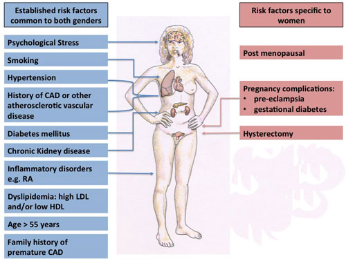
Typical risk factors should be considered during assessment of CVD risk and include, age > 55 years, pre-existing CAD, hypertension, dyslipidaemia and diabetes mellitus among others, see figure 1.
Risk factors specific to women include postmenopausal status, hysterectomy and complications during pregnancy. Women who develop gestational diabetes mellitus (GDM) or pre-eclampsia more than double their risk of developing CVD later in life.19
Physical inactivity is a significant risk factor for CVD. The Duke Activity Status Index (DASI) is a risk stratification tool measuring functional capacity and comprises a 12-item questionnaire. Women performing < 4.7 METs (metabolic equivalents) of effort in the form of activities of daily living were subject to a 3.7-fold increased risk of death or non-fatal MI when compared with women who engage in greater levels of activity.20
Menopausal status and CVD risk
Transition to the menopause is associated with a worsening CHD risk profile.21,22 After the menopause women may experience an increase in body weight, alteration in fat distribution, centripetal obesity and visceral fat deposition, with an associated increase in other CVD risk factors such as diabetes mellitus.23
Diabetes in particular is a potent risk factor in women, demonstrated by a meta-analysis of 37 studies and almost 450,000 patients which found a 50% greater relative risk of fatal CHD compared with men with the disease.24 This substantial gender difference in mortality is linked to a more adverse risk factor profile, smaller coronary vessel diameter and often inadequate treatment of diabetes in women.
As oestrogen production diminishes in aging women, systolic blood pressure tends to increase, modulated by the increase in plasma-renin activity.25-27 Additionally, salt sensitivity and sympathetic drive are increased in postmenopausal women.
Over the age of 75 isolated systolic hypertension is 14% more prevalent in women than men and a significant cause of morbidity due to left ventricular hypertrophy, diastolic heart failure and cerebrovascular disease.28
The risk of hypercholesterolaemia in younger women is lower compared with men. However, after the menopause women have a more adverse lipid profile, total cholesterol and low-density lipoprotein (LDL) levels increasing by 10% and 14% respectively without significant change in high-density lipoprotein
levels. 21,29 Postmenopausal re-evaluation of lipids may be an important consideration in women with a borderline premenopausal profile. Over the age of 65 mean LDL is higher in women compared with men but LDL reduction with statins has been shown to reduce CHD mortality to a similar extent.30
Gender differences in management
Underestimation of cardiovascular risk in women results in delayed or even missed diagnosis of CVD. Focusing on investigations which detect severe coronary stenosis is unlikely to yield accurate diagnoses as women have a lower prevalence of obstructive CAD but have greater symptom burden and functional impairment, see table 2. The management of stable angina in men and women remains similar, and includes lifestyle modifications, anti-anginal medications and pharmacological secondary prevention, revascularisation and rehabilitation.31
Cardiac investigations
The exercise ECG is of limited use in the evaluation of chest pain, owing to lower diagnostic accuracy for CAD in women compared with men (sensitivity and specificity around 60-70% vs 80% respectively).32,33 This is partly due to gender-related difficulties in reaching an adequate level of exercise (> 5 METs) required to diagnose inducible ischaemia.20 Hormonal variations may also contribute to false-positive rates in premenopausal women and hormone replacement therapy may cause false- negative results due to vasodilatory properties.34
Exercise stress echocardiography provides more definitive results than exercise ECG. However, pharmacological stress testing is also an accurate diagnostic technique and is preferred for diagnosing CAD in females with lower exercise capacity.35 Stress echocardiography using dobutamine has a sensitivity and specificity of 85% and 75% respectively for detecting CAD.10,20
Single photon emission computed tomography, a nuclear imaging technique used to evaluate stress-induced myocardial perfusion defects, is also highly accurate. However, image quality is affected by breast tissue and obesity which may result in false-positive results in women.
In the detection of obstructive CAD, CT coronary angiography has similar diagnostic accuracy in detecting ≥ 50% and ≥ 70% coronary stenosis in both sexes.36 Of note, in women over 55 years with intermediate risk for CHD, the lack of coronary calcium has a high negative predictive value (99%).37
Microvascular angina
This term refers to patients with symptoms of angina and abnormalities on stress testing, despite normal coronary arteries on angiography, with women more affected than men.
In a study of 886 patients presenting with chest pain and subsequently undergoing coronary angiography, women were five times as likely to have normal coronary arteries compared with men.38 However, the same risk factors causing atherosclerosis may cause microvascular disease and low oestrogen levels in women have also been implicated. Treatment is based on symptom control with calcium channel blockers and oral nitrates.
Stress cardiomyopathy
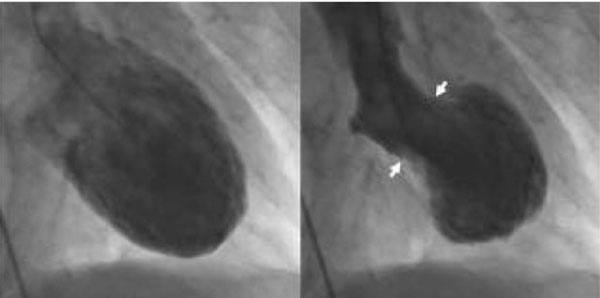
Stress (Takotsubo) cardiomyopathy is triggered by intense, unexpected emotional or physical stress and is characterised by transient apical systolic dysfunction or ballooning of the left ventricle, see figure 2. Presentation is usually similar to AMI with ST segment elevation on the ECG and raised cardiac troponin levels. However, there is no obstructive coronary disease and the observed regional wall motion abnormality is not limited to the territory supplied by a single coronary artery. The syndrome predominantly affects postmenopausal women and in a study of 1,750 patients from 26 centres, almost 90% were women with a mean age of 66.4 years.39
The pathogenesis is not fully understood but catecholamine toxicity, microvascular dysfunction and coronary artery spasm have all been highlighted as possible mechanisms. Familial cases raise the suspicion of a genetic component to predisposition, however more convincing is the finding that 56% of sufferers had a psychiatric or neurological disorder compared with 26% of patients with ACS.39
Gender-dependent outcomes
The Framingham Heart Study6 reported that women with angina had better outcomes and fewer adverse events e.g. AMI, than males. However, more recent data suggest that both men and women presenting with stable angina are subject to increased mortality when compared with the general population.40 Therefore stable angina is no longer viewed as a benign condition in women. Evidence to support this comes from the Euro Heart Survey of Stable Angina demonstrating that during the one year follow-up period, women had double the risk of death or non-fatal AMI compared with men.41
It seems that subgroups of CVD have varying outcomes. Women presenting with STEMI have worse outcomes compared with men.42 However, in those presenting with NSTEMI no differences in outcome were demonstrated.43
The pathogenesis of CVD may explain the differences in outcomes. Women with unstable angina are more likely to have non-obstructive CAD compared with men, and less extensive disease correlates with a better prognosis.7
In contrast, women presenting with STEMI, where the process of acute thrombus formation secondary to ruptured plaque results in total occlusion of their comparably smaller vessels, have a poorer prognosis.44
Women who present with AMI at a young age (< 50 years) have a worse prognosis compared with age-matched male counterparts.45 This is highlighted by a large study of more than 50,000 patients who underwent coronary artery bypass grafting between 1993 and 1999, a higher mortality rate was demonstrated among women under the age of 50 when compared with men (3.4% vs 1.1%).46 This trend tends to diminish with increasing age.
Improvements in the management of women
In the past, clinical trials have mainly recruited men making it difficult to determine any advantages specific to women. Despite similar recommendations for men and women, it has been well documented that diagnostic procedures and secondary prevention are not applied in women with the same intensity. Once a diagnosis of CVD is made the mainstay of treatment in women should be secondary prevention.
Medical management affects men and women in different ways and should therefore be tailored to gender.
For example, beta-blockers have been found to provide greater survival benefit in women with AMI than in men.47
The American Heart Association (AHA) guideline for the prevention of cardiovascular disease in women (2011) highlights the importance of lifestyle modification, smoking cessation and dietary intake in order to reduce morbidity and mortality secondary to CVD, see table 3.22 This guidance should inform national strategies for the prevention of CVD in women.
Further studies are required before sex specific guidelines can be robustly and reliably utilised.
Conclusion
There are significant differences in the presentation, response to treatment and outcomes between men and women.
In 2007, the annual mortality in women secondary to CAD was 4.7 times that of breast cancer.48 CVD in women continues to be a major public health concern owing to the difficulties facing physicians in identifying associated symptoms.
In addition, the general attitude that women are somehow protected against CVD compared with men makes the management of CVD in women all the more challenging. The degree of variation between the sexes is not yet known and whether these discrepancies are related to pathophysiology or merely differences in the detection of CVD is yet to be clarified. Ideally, an understanding of these variables will lead to gender specific prevention, identification and management of CVD in women with a beneficial effect on morbidity and mortality.
REFERENCES
1 Go AS, Mozaffarian D, Roger VL et al. Heart disease and stroke statistics-2014 update: a report from the American Heart Association. Circulation 2014;129:e28-e292
2 Roger VL, Weston SA, Gerber Y et al. Trends in incidence, severity, and outcome of hospitalized myocardial infarction. Circulation 2010;121(7):863-9
3 Towfighi A, Zheng L, Ovbiagele B. Sex-specific trends in midlife coronary heart disease risk and prevalence. Arch Intern Med 2009;169(19):1762-66
4 Douglas PS, Ginsburg GS. The evaluation of chest pain in women. N Engl J Med 1996;334:1311-15
5 Murabito JM, Evans JC, Larson MG et al. Prognosis after the onset of coronary heart disease. An investigation of differences in outcome between the sexes according to initial coronary disease presentation. Circulation 1993;88(6):2548-55
6 Lerner DJ, Kannel WB. Patterns of coronary heart disease morbidity and mortality in the sexes: A 26-year follow-up of the Framingham population. Am Heart J 1986;111(2):383-90
7 Hochman JS, Tamis JE, Thompson TD et al. Sex, clinical presentation, and outcome in patients with acute coronary syndromes. Global use of Strategies to Open Occluded Coronary Arteries in Acute Coronary Syndromes IIb Investigators. N Engl J Med 1999;341(4):226-32
8 Berger JS, Elliott L, Gallup D et al. Sex differences in mortality following acute coronary syndromes. JAMA 2009;302(8):874-82
9 Milner KA, Funk M, Arnold A et al. Typical symptoms are predictive of acute coronary syndromes in women. Am Heart J 2002;143(2):283-88
10 Shaw LJ, Bairey Merz CN et al. Insights from the NHLBI-sponsored Women’s Ischemia Syndrome Evaluation (WISE) study. Part I: Gender differences in traditional and novel risk factors, symptom evaluation, and gender-optimized diagnostic strategies. J Am Coll Cardiol 2006;47(3 Suppl):S4-S20
11 Rosengren A, Wallentin L, K Gitt A et al. Sex, age, and clinical presentation of acute coronary syndromes. Eur Heart J 2004;25(8):663-70
12 Diamond GA, Forrester JS, Hirsch M et al. Application of conditional probability analysis to the clinical diagnosis of coronary artery disease. J Clin Invest 1980;65(5):1210-21
13 Johnson BD, Shaw LJ, Pepine CJ et al. Persistent chest pain predicts cardiovascular events in women without obstructive coronary artery disease: Results from the NIH-NHLBI-sponsored Women’s Ischaemia Syndrome Evaluation (WISE) study. Eur Heart J 2006;27(12):1408-15
14 Canto JG, Rogers WJ, Goldberg RJ et al. Association of age and sex with myocardial infarction symptom presentation and in-hospital mortality. JAMA 2012;307: 813:813-22
15 Bugiardini R, Bairey Merz CN. Angina with “normal” coronary arteries: a changing philosophy. JAMA 2005;293(4):477-84
16 Grundtvig M, Hagen TP, German M et al. Sex-based differences in premature first myocardial infarction caused by smoking: twice as many years lost by women as by men. Eur J Cardiovasc Prev Rehabil 2009;16(2):174-79
17 Prescott E, Hippe M, Schnohr P et al. Smoking and risk of myocardial infarction in women and men: longitudinal population study. BMJ 1998;316(7137):1043-47
18 Vanhoutte PM, Shimokawa H, Tang EHC et al. Endothelial dysfunction and vascular disease. Acta Physiologica 2009;196:193-222
19 Heida KY, Franx A, Van Rijn BB et al. Earlier age of onset of chronic hypertension and type 2 diabetes mellitus after a hypertensive disorder of pregnancy or gestational diabetes mellitus. Hypertension 2015;66(6):1116-22
20 Shaw LJ, Olson MB, Kip K et al. The value of estimated functional capacity in estimating outcome: Results from the NHBLI-sponsored Women’s Ischemia Syndrome Evaluation (WISE) study. J Am Coll Cardiol 2006;47 (3 Suppl):S36-43
21 Matthews KA, Meilahn E, Kuller LH et al. Menopause and risk factors for coronary heart disease. N Engl J Med 1989;321(10):641-46
22 Mosca L, Benjamin EJ, Berra K et al. Effectiveness-based guidelines for the prevention of cardiovascular disease in women—2011 update: A Guideline from the American Heart Association. Circulation 2011;23(11):1243-62
23 Kip KE, Marroquin OC, Kelley DE et al. Clinical importance of obesity versus the metabolic syndrome in cardiovascular risk in women: A report from the Women’s Ischemia Syndrome Evaluation (WISE) Study. Circulation 2004;109(6):706-13
24 Huxley R, Barzi F, Woodward M. Excess risk of fatal coronary heart disease associated with diabetes in men and women: meta-analysis of 37 prospective cohort studies. BMJ 2006;332(7533):733-38
25 Lima R, Wofford M, Reckelhoff JF. Hypertension in postmenopausal women. Curr Hypertens Rep 2012;14(3):254-60
26 Dubey RK, Oparil S, Imthurn B et al. Sex hormones and hypertension. Cardiovasc Res 2002;53:688-708
27 Burt V, Whelton P, Roccella E et al. Prevalence of hypertension in the US adult population. Results from the Third National Health and Nutrition Examination Survey, 1988-1991. Hypertension 1995;25(3):305-13
28 Vasan RS, Larson MG, Leip EP et al. Impact of high-normal blood pressure on the risk of cardiovascular disease. N Engl J Med 2001;345(18):1291-97
29 Abbey M, Owen A, Suzakawa M et al. Effects of menopause and hormone replacement therapy on plasma lipids, lipoproteins and LDL-receptor activity. Maturitas 1999;33(3):259-69
30 Baigent C, Keech A, Kearney PM et al. Efficacy and safety of cholesterol-lowering treatment: prospective meta-analysis of data from 90,056 participants in 14 randomised trials of statins. Lancet 2005 366:1267-78 [erratum appears in Lancet 2005;366(9494):1358 and Lancet 2008;371(9630):2084]
31 Task A, Members F, Fox K et al. Guidelines on the management of stable angina pectoris: The Task Force on the Management of Stable Angina Pectoris of the European Society of Cardiology. Eur Heart J 2006;27(11): 1341-81
32 Hlatky MA, Pryor DB, Harrell FE et al. Factors affecting sensitivity and specificity of exercise electrocardiography. Multivariable analysis. Am J Med 1984;77(1):64-71
33 Kwok Y, Kim C, Grady D et al. Meta-analysis of exercise testing to detect coronary artery disease in women. Am J Cardiol 1999;83:660-6
34 Grzybowski A, Puchalski W, Zieba B et al. How to improve noninvasive coronary artery disease diagnostics in premenopausal women? Am Heart J 2016; 10;156(5): 964.e1–964.e5
35 Mulvagh SL, DeMaria AN, Feinstein SB et al. Contrast echocardiography: current and future applications. J Am Soc Echocardiog 2000;13:331-42
36 Budoff MJ, Dowe D, Jollis JG et al. Diagnostic performance of 64-multidetector row coronary computed tomographic angiography for evaluation of coronary artery stenosis in individuals without known coronary artery disease. J Am Coll Cardiol 2008;52(21): 1724-32
37 Raggi P, Gongora MC, Gopal A et al. Coronary artery calcium to predict all-cause mortality in elderly men and women. J Am Coll Cardiol 2008;52(1):17-23
38 Sullivan AK, Holdright DR, Wright CA et al. Chest pain in women: clinical, investigative, and prognostic features. BMJ 1994;308(6933):883-86
39 Templin C, Ghadri JR, Diekmann J et al. Clinical features and outcomes of Takotsubo (Stress) cardiomyopathy. N Engl J Med 2015;373(10):929-38
40 Hemingway H, McCallum A, Shipley M et al. Incidence and prognostic implications of stable angina pectoris among women and men. JAMA 2006;295(12):1404-11
41 Daly C, Clemens F, Lopez Sendon JL et al. Gender differences in the management and clinical outcome of stable angina. Circulation 2006;113(4):490-98
42 Wenger NK, Shaw LJ, Vaccarino V. Coronary heart disease in women: update 2008. Clin Pharmacol Ther 2008;83(1):37-51
43 Champney KP, Frederick PD, Bueno H et al. The joint contribution of sex, age and type of myocardial infarction on hospital mortality following acute myocardial infarction. Heart 2009;95(11):895-99
44 Fuster V. Elucidation of the role of plaque instability and rupture in acute coronary events. Am J Cardiol 1995;76:24C-33C
45 Vaccarino V, Parsons L, Peterson ED et al. Sex differences in mortality after acute myocardial infarction: changes from 1994 to 2006. Arch Intern Med ;169(19):1767-77
46 Vaccarino V, Abramson JL, Veledar E et al. Sex differences in hospital mortality after coronary artery bypass surgery: Evidence for a higher mortality in younger women. Circulation 2002;105(10):1176-81
47 ISIS Study Group. Randomised trial of intravenous atenolol among 16 027 cases of suspected acute myocardial infarction: ISIS-1. First International Study of Infarct Survival Collaborative Group. Lancet 1986;2(8498):57-66
48 Roger VL, Go AS, Lloyd-Jones DM et al. Heart disease and stroke statistics-2011 update: A report from the American Heart Association. Circulation 2011;123:e18-e209
ARTICLE IN PDF
 Download this article in PDF format
PRESET SEARCHES
PubMed
Women heart disease treatment differences
NICE evidence search
EXTERNAL WEBLINKS
We welcome your feedback
If you wish to comment on this article or have a question for the authors, write to: [email protected]