SEARCH

Bergin U, Lynch RM. Artefact mimicking torsades: treat the patient not the ECG. Practitioner 2017;261(1801):23-26
Artefact mimicking torsades: treat the patient not the ECG
22 Feb 2017
AUTHORS
Ms Una Bergin, Final Year Medical Student, University College Dublin, Ireland, Midland Regional Hospital, Mullingar, Co. Westmeath, Ireland
Dr Richard M Lynch MB BCh BAO MMedSc FRCSI FFAEM, Consultant in Emergency Medicine, Midland Regional Hospital, Mullingar, Co. Westmeath, Ireland, and Medway Maritime Hospital, Gillingham, Kent, UK
Article
Abstract
ECG interpretation is an essential skill in the management of the acutely unwell patient and in addition to history taking and physical examination has been shown to have a significant effect on referral patterns to cardiologists. One of the basic initial steps in ECG interpretation is assessment for the presence of artefact which if present can dramatically influence the diagnosis. The most common sources of artefact are tremor e.g. in Parkinson’s disease, loose skin electrodes and electromagnetic interference from other medical devices and mobile phones. The Medicines and Healthcare products Regulatory Agency in the UK advises that mobile phones should be kept at least one metre away from equipment that is sensitive to electromagnetic interference. The possibility that artefact is the cause of the ECG appearance should always be considered if bizarre ECG changes are present particularly in an asymptomatic patient. The ECG should always be interpreted in the context of the patient’s condition. If artefact is thought to be the cause of the ECG appearance, then any contributory factors present should be corrected and the ECG should be repeated before invasive investigations are undertaken or treatment is administered.
Article
The ECG is invaluable in the assessment and management of the acutely unwell patient and is one of the core competencies for training in general practice1 and emergency medicine.2
It is easy to perform, reproducible, cheap and when combined with patients’ symptoms is highly specific for time critical conditions such as myocardial infarction (MI).3
In recent years, in the UK and Ireland, there has been a marked increase in the availability and use of ECG machines in general practice. In 2008, Day and colleagues reported that 85% of GPs who responded to their survey had an ECG machine and that 91% of them used it at least once a week.4 In two earlier studies only 11%5 and 43%6 of GP respondents respectively stated that they routinely performed an ECG on patients with a suspected MI.
We report a case in which artefact was misinterpreted as torsades de pointes, often referred to simply as torsades. Our patient did not suffer any harm and did not have any unnecessary investigations, yet inappropriate treatment was administered making the potential for harm a real possibility.
We describe the ECG features which differentiate artefact from torsades and review common sources of ECG artefact, in both hospital and general practice.
Case report
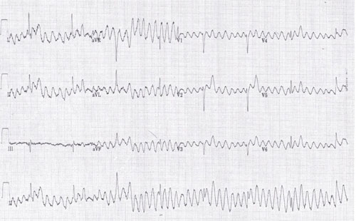
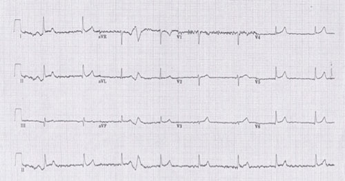
A 61-year-old man presented to our emergency department following a witnessed presyncopal episode.There was no associated chest pain, dyspnoea or loss of consciousness. His medical history revealed that he had suffered a stroke a few years earlier, from which he had made a complete recovery. Vital signs were within normal limits. No abnormality was observed on clinical examination. An ECG was obtained on presentation and was interpreted as torsades de pointes, see figure 1. He was treated with 2 g magnesium sulphate which was administered intravenously. A second ECG was recorded 15 minutes later and it showed complete resolution of the abnormal ECG pattern, see figure 2.
Discussion
ECG interpretation is an essential skill in the management of the acutely unwell patient and in addition to history taking and physical examination has been shown to have a significant effect on referral patterns to cardiologists.7 Rutten and colleagues observed a 34% reduction in specialist referrals among patients who previously would have been referred and a 20% increase in referrals among those who previously would not have been referred. Medical management was altered in 40% of patients.7
The conundrum we faced was deciding whether the ECG appearances represented torsades or artefact resulting in a spurious diagnosis of torsades. ECG features consistent with torsades included rapid, irregular, broad QRS complexes of different sizes and shapes and the apparent twisting of these complexes around the isoelectric line, particularly seen in the rhythm strip, see figure 1.8
All these ECG changes resolved completely within 15 minutes of administration of magnesium, see figure 2. In an early study into the effectiveness of intravenous magnesium in the management of torsades, Tzivoni and colleagues reported that in 9 out of 12 patients with torsades the tachyarrhythmia completely resolved within 1-5 minutes of administration of magnesium. The other three resolved after treatment with a second dose of magnesium given 5-15 minutes after the first dose.9
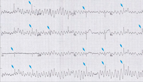
In lead III, see figure 3 there is no visible evidence of torsades, which should be present in all leads, and QRS complexes are clearly identifiable within the abnormal rhythm in most of the other leads. Both these factors confirm that the ECG appearances are due to artefact mimicking torsades.10
Torsades arises due to prolongation of the QT interval and is associated with an increased risk of developing ventricular fibrillation and subsequently sudden death.11 Urgent intervention is required when diagnosed.
The most common causes of prolonged QT interval are medications such as amiodarone and erythromycin, structural cardiac disease and electrolyte abnormalities, especially hypokalaemia and hypomagnesaemia.11
Conversely, misdiagnosing artefact as torsades can lead to unnecessary investigations, interventions and cost, with the possibility of significant complications due to unnecessary treatments, examples of which have included inappropriate defibrillation,12 insertion of a permanent pacemaker, and internal defibrillator for a broad complex tachyarrhythmia which was later identified as artefact.13
Unfortunately, we did not have any previously recorded ECGs for assessment of the QT interval, although a normal ECG may have inadvertently added weight to the possible diagnosis of torsades.
Assessing for artefacts
One of the basic initial steps in ECG interpretation is assessment for the presence of artefact, which if present, can dramatically influence the diagnosis.12,14,15
The most common sources of artefact are tremor e.g. in Parkinson’s disease,14,16 loose skin electrodes17 and electromagnetic interference from other medical devices and mobile phones.13,18
Unfortunately, doctors do not always recognise when the ECG appearances are due to artefact as highlighted by this case. Knight and colleagues showed an ECG, with artefact resembling a broad complex tachycardia, to 766 doctors.19
Only 6% (3 of 55) of interns, 42% (93 of 221) of cardiologists and 62% (304 of 490) of electrophysiologists correctly identified artefact as the cause of the broad complex tachycardia.
Of those who failed to identify the rhythm correctly 31% of internists, 53% of cardiologists and 88% of electrophysiologists stated that they would recommend invasive procedures for further investigation and management of the broad complex tachycardia.19
A wide range of commonly available healthcare electrical equipment has been reported to interfere with the ECG recording.20 Periyasamy and Dhanasekaran investigated the effect that electromagnetic interference from 2G and 3G mobile phones had on a variety of medical equipment when placed in close proximity to them.21
The ECG tracing became distorted during the ringing or conversation modes. Defibrillators were not susceptible to electromagnetic interference from mobile phones.
The authors have not quantified the magnitude of this effect and have not reported if the artefact produced was clinically relevant.21 The distance that mobile phones should be kept away from equipment sensitive to electro-magnetic interference varies from as little as 7.5 cm22 to as much as 1.25 m.23
The Medicines and Healthcare products Regulatory Agency in the UK advises that mobile phones should be kept at least one metre away from equipment sensitive to electromagnetic interference but they do not state how this recommendation was reached.24
There is a consensus that the highest risk of electromagnetic interference occurs with the use of two-way radios and mobile phones.25 Ishihara and colleagues studied the effect that third generation mobile phones had on 32 commonly available medical devices.26 Twelve medical devices (37.5%) were affected by electromagnetic interference which was shown to be dependent on the transmission power and the distance between the mobile phone and the medical device.26
However, Tri and colleagues conducted 300 tests with two different mobile phones in 75 patients’ rooms.
No interference was detected.27
Despite the serious complications, which can arise if ECG changes due to artefact are not recognised, ECG interpretation books largely fail to highlight this problem. There are, however, some notable exceptions.28
The possibility that artefact is the cause of the ECG appearance should always be considered if bizarre ECG changes are present particularly in an asymptomatic patient.
The ECG should always be interpreted in the context of the patient’s condition. If artefact is thought to be the cause of the ECG appearance, then any contributory factors present should be corrected and the ECG should be repeated before invasive investigations are undertaken or treatment given.16
Conclusion
It is important to consider artefact as the cause of bizarre ECG appearances. While it might not always be possible to identify the cause of the artefact it is important for GPs and hospital clinicians to recognise the presence of artefact.
If the ECG changes are thought to be due to artefact the ECG should be repeated before investigations or treatment are initiated. Common sources of electromagnetic interference should be identified and corrected.
REFERENCES
1 The RCGP Curriculum: Professional & clinical modules. 2.01–3.21 Curriculum modules, Version approved 19 January 2016 for implementation from 1 February 2016
2 The Royal College of Emergency Medicine. Curriculum and assessment systems for training in emergency medicine. August 2015. Curriculum revised and applicable from August 2016. Approved 23 November 2015
3 Herring N, Paterson DJ. ECG diagnosis of acute ischaemia and infarction: past, present and future. QJM 2006;99:219-30
4 Day A, Oldroyd C, Godfrey S, Quinn T. Availability of cardiac equipment in general practice premises in a cardiac network: a survey. Br J Cardiol 2008;15:141-44
5 Macallan DC, Bell JA, Braddick M et al. The electrocardiogram in general practice: its use and its interpretation. J R Soc Med 1990;83:559-62
6 Murphy AW, Power R, Ungruh K, Bury G. Pre-hospital management of acute myocardial infarction. Ir J Med Sci 1992;161:593-96
7 Rutten FH, Kessels RGH, Willems FF, Hoes AW. Electrocardiography in primary care; is it useful? Int J Cardiol 2000;74(2-3):199-205
8 Drew BJ, Ackerman MJ, Funk M et al. Prevention of torsade de pointes in hospital settings: A Scientific Statement from the American Heart Association and the American College of Cardiology Foundation. Circulation 2010;121(8):1047-60
9 Tzivoni D, Banai S, Schuger C et al. Treatment of torsade de pointes with magnesium sulfate. Circulation 1988;77(2):392-97
10 Lin SL, Wang SP, Kong CW, Chang MS. Artefact simulating ventricular and atrial arrhythmia. Jpn Heart J 1991;32:847-51
11 Roden DM. Torsades de Pointes. Clin Cardiol 1993;16:683-86
12 Knight BP, Pelosi F, Michaud GF et al. Clinical consequences of electrocardiographic artefact mimicking ventricular tachycardia. N Engl J Med 1999;341(17):1270-74
13 Patadia M, Bartindale M, Caballero N. Recurrent asystole as an electrocardiographic artefact during microdebrider use in functional endoscopic sinus surgery, Am J Rhinol Allergy 2015;29(6):e220-e223
14 Chase C, Brady WJ. Artefactual electrocardiographic change mimicking clinical abnormality on the ECG. Am J Emerg Med 2000;18(3):312-16
15 Llinas R, Henderson GV. Images in clinical medicine. Tremor as a cause of pseudo-ventricular tachycardia. N Engl J Med 1999;341(17):1275
16 Srikureja W, Darbar D, Reeder GS. Tremor-induced ECG artefact mimicking ventricular tachycardia. Circulation 2000;102:1337-38
17 Davis-Smith C. Skin preparation to reduce ECG artefact. Biomed Instrum Technol 2000;34(4):246
18 Mariappan PM, Raghavan R, Abdel Aleem SHE, Zobaa AF. Effects of electromagnetic interference on the functional usage of medical equipment by 2G/3G/4G cellular phones: A review. J Adv Research 2016;7:727-38
19 Patel SI, Souter MJ. Equipment-related electrocardiographic artefacts causes, characteristics, consequences, and correction. Anesthesiol 2008;108:138-48
20 Knight BP, Pelosi F, Michaud GF et al. Physician interpretation of electrocardiographic artefact that mimics ventricular tachycardia. Am J Med 2001;110:33-35-8
21 Periyasamy M, Dhanasekaran R. Evaluation of electromagnetic interference between critical medical devices and new generation cellular phones. J Microw Power Electromagn Energy 2015;3:160-70
22 Buczkowski T, Janusek D, Zavala-Fernandez H et al. Influence of mobile phones on the quality of ECG signal acquired by medical devices. Measurement Science Review 2013;13(5):231-36
23 Helhel S, Colak ZA, Ozen S. Distance and location of both mobile phones and health care units: Determines the interference level. Am J Biomed Engin 2011;1(2):78-82
24 www.gov.uk/government/publications/
electromagnetic-interference-sources/electromagnetic-interference-sources#mobile-phones
25 Boyle J. Wireless technologies and patient safety in hospitals. Telemed J E Health 2006;12(3):373-82
26 Ishihara S, Higashiyama J, Onishi T. Electromagnetic interference with medical devices from third generation phone including LTE. Electromagnetic Compatibility, Tokyo (EMC'14/Tokyo), 2014 International Symposium on. IEEE, 2014
27 Tri JL, Severson RP, Hyberger LK, Hayes DL. Use of cellular telephones in the hospital environment. Mayo Clin Proceed 2007;82(3):282-85
28 Crawford J, Doherty L. Practical aspects of ECG recording. M & K Publishing. Keswick, UK. 2012. ISBN 978-1-905533930-7
FURTHER READING
FURTHER READING
Practical aspects of ECG recording By L Doherty and J Crawford, M & K Publishing. Keswick, UK. 2012 ISBN 978-1-905533930-7
ARTICLE IN PDF
 Download this article in PDF format
CLICK ON IMAGES
PRESET SEARCHES
PubMed
ECG artefact mimicking torsades
Immediate CPD prompts
These handy templates*, associated with all articles, prompt drafting of personal learning, reflection and planning.
Save your record straight into your personal development folder on your device or cloud storage system.
*Adobe fillable PDFs. Adobe reader needed for iPad
Comment or question
If you, or your peer group, would like to comment on an article or have a question for the authors, write to:
Editor
Case report submissions
Do you have an interesting case that you would like to share with other readers?
Your article would need to be around 1,500 words long, include a brief review of the literature to put the case in context, and be fully referenced in the Vancouver style.
Submissions for consideration for publication should be sent to: [email protected]