SEARCH

Moßhammer D, Hlobil H, Joos S, Reichert W. A patient with Lyme arthritis presenting in general practice. Practitioner 2013;257 (1758): 25-27
A patient with Lyme arthritis presenting in general practice
21 Feb 2013
AUTHORS
Dr Dirk Moßhammer 1, 2 MD MPH
Dr Harald Hlobil3 MD
Dr Stefanie Joos4,5 MD
Dr Walter Reichert 2 MD
1 University Hospital Tübingen, Division of General Practice, Tübingen, Germany
2 Practice of Family Medicine and Internal Medicine, Nufringen, Germany
3 Professional Partnership Practice of Laboratory Medicine, Sindelfingen, Germany
4 Department of General Practice and Health Services Research, University Hospital Heidelberg, Heidelberg, Germany
5 Competence Centre General Practice Baden-Württemberg, University Hospital Heidelberg, Heidelberg, Germany
Article
Lyme disease, also known as Lyme borreliosis, is the most common tick-borne infectious disease in Europe and North America. Infection in humans often takes place in woodland or heath areas where animals that carry the ticks such as deer and mice are found.
The Health Protection Agency estimates that there are 2,000 to 3,000 cases of Lyme disease in England and Wales each year (see box 1) and that around 15% of infections are acquired abroad.1 It is caused by infection with Borrelia burgdorferi sensu lato (B. burgdorferi s.l.) complex. Borrelia is a Gram-negative spirochaete bacterium.
In Europe B. burgdorferi sensu stricto, B. afzelii, B. garinii, B. bavariensis and B. spielmanii can cause Lyme disease, whereas in the USA B. burgdorferi sensu stricto is the sole culprit.2
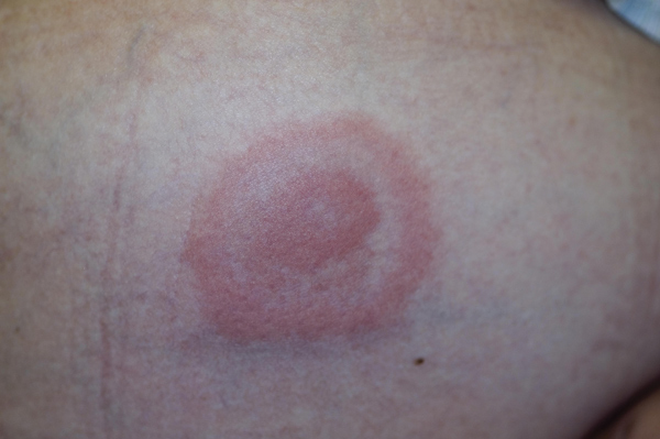
In principle, Lyme disease may affect every organ of the body and may manifest in different stages. Early localised or disseminated stages are characterised by erythema migrans (see figure 1), lymphadenosis benigna cutis, facial palsy and arthritis and the later stages by arthritis, acrodermatitis chronica atrophicans or encephalomyelitis.3 Not all these features occur in every case. The incubation time of the earlier stages varies from several days to months, and that of the later stages from weeks to months, and even years.4
Lyme arthritis commonly manifests mono- or oligoarticularly (< 5 joints). Most frequently the knee joint is affected, followed by the ankle, the wrist and the elbow.5
According to epidemiology data, there were around 16,500 reported cases of Lyme disease in Germany from 2007 to 2009. The presence of Lyme arthritis in Lyme disease cases ranges from around 2% to 8%.4, 6 However, a figure of almost 25% was reported by primary care physicians in a nationwide survey in Germany.7
A prospective US study from the 1980s suggests that not treating erythema migrans with antibiotics results in an increase in Lyme arthritis. During the six-year follow-up, more than half of the 55 patients included with erythema migrans suffered from Lyme arthritis.5
The work-up of Lyme arthritis should include a careful history including residence in, or time spent visiting, an endemic region, previous history of tick bite(s), and erythema migrans. Clinical examination for arthritis, serology and other laboratory investigations are also key, see below. As immunity after infection with B. burgdorferi s.l. cannot necessarily be assumed, reinfections are possible.8 We report a case history of a patient with Lyme arthritis presenting in a family practice in Nufringen, Germany.
Case history
A 63-year old woman with no pre-existing disorders presented with pain in her left knee, which had begun about two weeks before her first visit. She reported that redness, warmth and swelling of the knee had progressed markedly over the past three days. She also complained of general discomfort and feeling cold.
She had developed two red marks on her tibial skin, each about 2 cm in diameter, several weeks before the knee pain began. The marks were itchy and disappeared after a week. She recalled having a tick bite about three years ago and previous tick bites when she was a young woman. She was taking no medications and had no family history of gout, arthritis or rheumatic diseases.
Physical findings
The patient was only able to walk with the aid of crutches. There was marked swelling and warmth in the left knee, reddening of the distal ventro-lateral thigh and discrete swelling of the popliteal region and calf without reddening. She felt no pain on palpation of the calf. There was a large effusion of the knee joint and no knee flexion was possible.
Examination of the abdomen, skin and lymphatic system, heart, lungs and oral mucosa were normal. She had no fever. Blood pressure was 130/70 mmHg and her pulse regular.
Investigations
Duplex ultrasonography of the deep veins of the legs excluded deep vein thrombosis. Electrocardiogram and X-ray of the knee were normal.
Histology of the synovial biopsy material revealed a marked chronic synovitis with inflammatory phlegmon that was consistent with septic arthritis.
Borrelia was detected by PCR and DNA sequencing of the synovial fluid, and by serology follow-up of Borrelia-induced immune reactions.
The serology findings showed specific IgG antibodies against B. burgdorferi s.l. The detection of B. burgdorferi DNA by means of PCR and DNA sequencing of the synovial fluid confirmed the diagnosis of Lyme arthritis caused by B. garinii. Specific IgM antibodies could not be detected. Key findings are summarised in table 1.
Treatment
At the first visit to the practice, about 50 ml of unclear serous fluid was aspirated from the knee joint and taken to the laboratory immediately. The patient received oral ibuprofen (600 mg tid), and was instructed to cool her knee until the next day when the first laboratory results were expected.
As the cell count of the synovial fluid was 21,200/µl with a high proportion of segmented granulocytes the patient was admitted to hospital for knee lavage because a septic arthritis was suspected. In hospital she also received analgesic and antibiotic therapy and physiotherapy for the knee joint. Antibiotic treatment included cefazoline IV for one week followed by oral cefadroxil (1g bid).
For thromboprophylaxis, she received LMW heparin subcutaneously.
After 11 days, she was discharged on cefadroxil. Then, back in the community, the antibiotic therapy was continued with ceftriaxone (2 g/day) IV.
After 14 weeks the patient had completely recovered.
Discussion
Besides the common causes of acute and subacute gonarthritis (i.e. gouty and septic arthritis, activated osteoarthritis and rheumatologic disorders), Lyme arthritis needs to be considered in patients who live in, or have visited, endemic regions. The lack of IgM antibodies and the confirmation of IgG antibodies raised the suspicion of Lyme arthritis.
Synovial fluid/synovial biopsy material
The cytology of the synovial fluid (granulocytes were prevailing) and the histology of the synovial membrane (marked chronic synovitis with inflammatory phlegmon) were consistent with the findings in septic arthritis. However, bacterial growth could not be detected.
The histology findings in Lyme synovitis are associated with an unspecific inflammatory reaction that can also be found in posttraumatic synovitis, rheumatic arthritis and, juvenile idiopathic arthritis.9,10
The detection of spirochaete bacteria in the biopsy material is likely to be successful in only a few cases. This procedure is time consuming and work intensive, therefore, it is not recommended for the routine diagnostic work-up in Lyme arthritis.11
Rather, diagnosis of Lyme arthritis can be supported by PCR and DNA sequencing of the synovial fluid or synovial biopsy material.12 The sensitivity of the detection of DNA is about 50-70%; DNA can more often be detected in the synovial membrane than in the synovial fluid.13 A negative PCR test result does not exclude Lyme arthritis, whereas a positive result practically confirms it.13
In our patient, DNA of B. garinii could be detected.
Serology findings
In order to confirm the diagnosis of Lyme arthritis clinical findings and specific IgG antibodies are necessary. The lack of IgG antibodies practically rules out Lyme arthritis.11,14,15,16
At the first visit the patient had high blood concentrations of antibodies (by ELISA), after five weeks the parallel testing showed an increase and after 14 weeks a decline. The patient had no IgM antibodies, however IgM antibodies are often not detected in Lyme arthritis.17, 18,19 Several antibodies against recombinant antigens of B. burgdorferi s.l. could be detected by Luminex technology (see box 2).
Therapy
The patient initially, in hospital, received a first-generation cephalosporin. The recommendations from the European Concerted Action on Lyme Borreliosis (EUCALB), however, include second- and third- generation cephalosporins. Therefore, we decided to administer ceftriaxone (2 g/day, IV, for 4 weeks).
The EUCALB therapy recommendations for adults are as follows:16
• Tetracycline antibiotic of choice is doxycycline (100 mg bid, except for children under the age of eight years, pregnant women and breastfeeding mothers) or
• Aminopenicillin of choice is oral amoxicillin (500-1,000 mg tid) or
• A third-generation cephalosporin (explicitly mentioned is ceftriaxone, IV, 2 g/day; alternatively cefotaxime, IV, an exact dose is not specified).
For the latter, Nau and colleagues alternatively recommend cefuroxime, a second-generation cephalosporin (500 mg bid).20
In general, the prognosis of Lyme disease is assumed to be good, in particular after antibiotic therapy for early manifestations. However, clinical follow-up data after antibiotic therapy in Lyme arthritis are sparse.20,21
Conclusion
The diagnosis of Lyme arthritis may be complex, especially the interpretation of serology data. As a rule, IgM antibodies are generally not detected in Lyme arthritis, whereas IgG antibodies are mandatory for diagnosis. Antibodies can be detected even years after infection(s) in asymptomatic persons with previous Lyme disease treated with antibiotics. If an assessment of serology follow-up data is considered, at least parallel testing of the sera has to be performed. However, such blood samples need to be stored deep frozen, and in practice this can be rarely managed.
Arthralgia with IgG antibodies persistent from previous infection(s) needs to be distinguished from Lyme arthritis. DNA analysis is a valuable additional method to diagnose Lyme arthritis.
After infection, immunity against B. burgdorferi sensu lato cannot necessarily be assumed. Hence, reinfections are possible.
In contrast to the USA, one has to keep in mind that in Europe infections with different endemic species of the B. burgdorferi sensu lato complex occur.
Acknowledgement
The authors would like to thank Dr Rainer Oehme, Board of Health of the Federal State Baden-Württemberg, Stuttgart, Germany for DNA sequencing of the synovial fluid.
REFERENCES
1 www.hpa.org.uk/Topics/ InfectiousDiseases/ InfectionsAZ/ LymeDisease/ EpidemiologicalData/lymLymeepidemiology/
2 Fingerle V. Lyme-Borreliose: Wissenswertes zum Vektor und die mikrobiologische Diagnostik. Der Mikrobiologe 2011;3(21):83-92
3 Satz N. Klinik der Lyme-Borreliose. Hans Huber. Bern. 2010
4 RKI. Lyme-Borreliose: Analyse der gemeldeten Erkrankungsfälle der Jahre 2007 bis 2009 aus den sechs östlichen Bundesländern. Epidemiologisches Bulletin 2010;12:101-10
5 Steere AC, Schoen RT, Taylor E. The clinical evolution of Lyme arthritis. Ann Intern Med 1987;107(5):725-31
6 Schmidt R, Kabatzki J, Hartung S, Ackermann R. Erythema migrans borreliosis in the Federal Republic of Germany. Epidemiology and clinical aspects. Dtsch Med Wochenschr 1985;110(47):1803-7
7 Priem S, Munkelt K, Franz JK et al. Epidemiology and therapy of Lyme arthritis and other manifestations of Lyme borreliosis in Germany: results of a nation-wide survey. Z Rheumatol 2003;62(5):450-8
8 Krause PJ, Foley DT, Burke GS et al. Reinfection and relapse in early Lyme disease. Am J Trop Med Hyg 2006;75(6):1090-4
9 Duray PH. Histopathology of clinical phases of human Lyme disease. Rheum Dis Clin North Am 1989;15(4):691-710
10 Duray PH, Steere AC. Clinical pathologic correlations of Lyme disease by stage. Ann N Y Acad Sci 1988;539:65-79
11 Brouqui P, Bacellar F, Baranton G et al. Guidelines for the diagnosis of tick-borne bacterial diseases in Europe. Clin Microbiol Infect 2004;10(12):1108-32
12 Steere AC, McHugh G, Damle N, Sikand VK. Prospective study of serologic tests for lyme disease. Clin Infect Dis 2008;47(2):188-95
13 Aguero-Rosenfeld ME, Wang G, Schwartz I, Wormser GP. Diagnosis of lyme borreliosis. Clin Microbiol Rev 2005;18(3):484-509
14 O'Connell S. Lyme borreliosis: current issues in diagnosis and management. Curr Opin Infect Dis 2010;23(3):231-5
15 Strle F, Stanek G. Clinical manifestations and diagnosis of lyme borreliosis. Curr Probl Dermatol 2009;37:51-110
16 www.eucalb.com
17 Akin E, McHugh GL, Flavell RA et al. The immunoglobulin (IgG) antibody response to OspA and OspB correlates with severe and prolonged Lyme arthritis and the IgG response to P35 correlates with mild and brief arthritis. Infect Immun 1999;67(1):173-81
18 Craft JE, Grodzicki RL, Shrestha M et al. The antibody response in Lyme disease. Yale J Biol Med 1984;57(4):561-5
19 Wilske B, Fingerle V, Schulte-Spechtel U. Microbiological and serological diagnosis of Lyme borreliosis. FEMS Immunol Med Microbiol 2007;49(1):13-21
20 Nau R, Christen HJ, Eiffert H. Lyme disease-current state of knowledge. Dtsch Arztebl Int 2009;106(5):72-81
21 Kalish RA, Kaplan RF, Taylor E et al. Evaluation of study patients with Lyme disease, 10-20-year follow-up. J Infect Dis 2001;183(3):453-60
ARTICLE IN PDF
 Download this article in PDF format