SEARCH
 = Registered users
 = Paid-up subscribers
Newell S, Overton C. Postmenopausal bleeding should be referred urgently. Practitioner 2012;256 (1749):13-15
Postmenopausal bleeding should be referred urgently
21 Mar 2012
AUTHORS
Dr Sarah Newell MBBS, Specialty trainee in Obstetrics and Gynaecology
Mrs Caroline Overton MBBS MD FRCOG FHEA, Consultant Obstetrician & Gynaecologist, Subspecialist in Reproductive Medicine & Laparoscopic Surgery
St Michael's University Hospital, Bristol
Article
Postmenopausal bleeding is an episode of bleeding 12 months or more after the last menstrual period.1 It is one of the most common reasons for referral to the gynaecology department. All women with postmenopausal bleeding should be referred urgently, endometrial cancer is present in approximately 10% of cases.1
Causes
Postmenopausal bleeding is a common problem and occurs in up to 10% of women aged over 55 years. The majority of cases have a benign cause. There is no evidence to indicate whether different patterns of postmenopausal bleeding such as one-off bleeding or more frequent bleeds are more likely to be associated with malignancy.1
Possible causes of postmenopausal vaginal bleeding are listed in table 1.
Primary care assessment
The aim of assessment and investigation of postmenopausal bleeding is to identify a cause and exclude cancer. Assessment should start by taking a detailed history with identification of risk factors for endometrial cancer (see table 2) as well as a medication history covering use of HRT, tamoxifen and anticoagulants.
Abdominal and pelvic examinations should be carried out to look for masses. Speculum examination should be performed to:
• see if a source of bleeding can be identified
• assess atrophic changes in the vagina
• look for evidence of cervical malignancy or polyps.
The woman is usually clear where the bleeding has come from i.e. from the vagina, urethra or rectum. When there is uncertainty about the origin of the bleeding a tampon can be inserted to confirm the bleeding is vaginal rather than rectal or urethral.
If a source of bleeding is identified on speculum, treatment for this should be initiated. The woman should have an ultrasound scan arranged to check the endometrial thickness. If the endometrial thickness is be taken.
Diagnosis
All women who have an episode of postmenopausal bleeding should be seen under the two-week referral rule. Endometrial cancer should be excluded.
Ultrasound scan and endometrial biopsy are complementary. Ultrasound scan can define endometrial thickness and identify structural abnormalities of the uterus, endometrium and ovaries. Endometrial biopsy provides a histological diagnosis.
Transvaginal ultrasound scan
Most evidence at present advocates the use of transvaginal ultrasound scan (TVUS) as the initial investigation of postmenopausal bleeding. TVUS can reliably assess the thickness of the endometrium and identify structural abnormalities such as polyps or submucous fibroids. It is also a valuable diagnostic tool in excluding ovarian malignancy.
The measurement of endometrial thickness aims to identify which women with postmenopausal bleeding are at significant risk of endometrial cancer. The thicker the endometrium, the higher the chance of endometrial cancer being present. The chance of finding endometrial cancer in a woman with an endometrial thickness of ≤ 4 mm is 0.8%.4
The thinner the endometrial thickness chosen as a cut-off, the fewer cases of endometrial cancer will be missed. A higher cut-off will result in more cases of endometrial cancer being missed but will mean fewer unnecessary investigations.
The SIGN guidelines1 advise that an endometrial thickness of of < 3 mm can be used to exclude endometrial cancer in women who:
• have never received HRT
• have not used HRT for a year or more
• are on continuous combined HRT
A recent meta-analysis suggested that a cut-off of 5 mm was a reasonable compromise.9 It is important to remember that no endometrial thickness completely excludes cancer and the disease can present in women without postmenopausal bleeding.
If the examination is normal, the bleeding has stopped and the endometrial thickness is < 5 mm on TVUS, no further action need be taken. If bleeding recurs, referral for hysteroscopy would be indicated.5
Endometrial biopsy
Endometrial sampling is an effective screening test for endometrial cancer but misses benign structural abnormalities such as endometrial polyps. Blind endometrial biopsy techniques can be used to obtain samples. Endometrial samplers use negative pressure to allow aspiration of tissue. All blind sampling of the endometrium will miss some cancers. Outpatient endometrial sampling will fail to obtain a sample in approximately 7% of cases.8
Further investigations
Hysteroscopy and curettage is the gold standard investigation as it allows direct visualisation and assessment of the uterine cavity and any structural abnormalities, and directed biopsy of specific lesions. Hysteroscopy is indicated in cases in which endometrial sampling cannot be performed because of cervical stenosis or discomfort; or where bleeding persists after negative biopsy.
If the endometrium is > 5 mm in thickness or the scan has identified a structural abnormality such as a polyp or submucous fibroid then further investigation is justified.
One stop clinics
Recent innovations include the development of specialist one stop clinics which enable clinical examination, ultrasound scan, endometrial sampling and outpatient hysteroscopy to be performed at the same visit. Following this assessment, reassurance can be offered or further investigations and treatment can be arranged.
Management
Benign conditions
Vaginal atrophy can be treated using topical oestrogens and there is minimal systemic absorption. Our preferred approach is to use vaginal oestrogen daily for two weeks and then once or twice weekly for maintenance. This can be continued long term without the need for any monitoring. If other menopausal symptoms are present then HRT may be used.
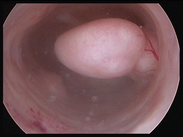
Cervical polyps can usually be removed easily as an outpatient procedure. Sponge forceps are used to twist the pedicle gradually until the polyp comes away. Endometrial polyps should be removed hysteroscopically.
Endometrial hyperplasia without atypia has a good response rate when treated with progestagens. Complex endometrial hyperplasia with atypia progresses to endometrial carcinoma in 23% of cases5 and should therefore be treated as endometrial cancer.
Endometrial cancer
The treatment for endometrial cancer is hysterectomy. Endometrial cancer is staged using the FIGO staging system, and treatment is determined by the stage of the cancer.
Stage 1 (cancer confined to the uterus) is treated with hysterectomy and bilateral salpingoophorectomy. Stage 2 or high-risk stage 1 cancer requires lymph node dissection. Adjuvant therapy (usually radiotherapy) with or without chemotherapy may be offered depending on the final histological diagnosis and staging.
Conclusion
Postmenopausal bleeding is a relatively common presentation that usually has a benign cause. Endometrial cancer is present in approximately 10% of cases. All women with postmenopausal bleeding should be referred to the gynaecology department under the two week rule. First-line investigation is a TVUS, which provides valuable information to identify which women need to undergo more extensive investigations. A normal TVUS is reassuring, and if examination is normal further investigation is not required, providing the bleeding has stopped.
REFERENCES
1 Scottish Intercollegiate Guidelines Network. SIGN 61. Investigation of postmenopausal bleeding. SIGN. Edinburgh. 2002
2 Uterine Cancer (C54-C55) Average number of new cases per year and age-specific incidence rates, UK 2006-2008. info.cancerresearchuk.org/cancerstats/types/uterus/incidence/
3 Palmer J, Perunovic B, Tidy J. Endometrial hyperplasia. The Obstetrician & Gynaecologist 2008;10:211–216
4 Munot S, Lane G. Modern management of postmenopausal bleeding. Trends Urol Gynaecol Sex Health 2008;13:20-24
5 Montgomery B, Daum G, Dunton C. Endometrial Hyperplasia: A Review. Obstet Gynaecol Surv 2004;59:368-378
6 Olt G, Mortel R. Hormone-producing tumors of the ovary. Endocr Relat Cancer 1997;4:447-457
7 Parkin D. Cancers attributable to reproductive factors in the UK in 2010. Br J Cancer 2011;105:(S2)S73-S76
8 Clark T, Mann C, Shah N et al. Accuracy of outpatient endometrial biopsy in the diagnosis of endometrial cancer: a systematic quantitative review. Br J Obstet Gynaecol 2002;109:313-321
9 Gupta J, Chien P, Voit D et al. Ultrasonographic endometrial thickness for diagnosing endometrial pathology in women with postmenopausal bleeding: a meta-analysis. Acta Obstet Gynecol Scandinav 2002:81;799–816
ARTICLE IN PDF
EXTERNAL WEBLINKS
PDQ® (Physician Data Query) is NCI's cancer database of peer-reviewed, regularly updated evidence-based, referenced summaries
Endometrial screening
Endometrial
PubMed
NICE Evidence - selected by NICE
PubMed
Premature menopause hormone replacement
NICE Evidence
British National Formulary
Treatment of vaginal and vulval conditions
Current evidence - Ovarian cancer
US NCI
PDQ® (Physician Data Query) is NCI's cancer database of peer-reviewed, regularly updated evidence-based, referenced summaries
Ovarian
PubMed
UK NICE
Ovarian
UK Risk Assessment Tools
QCancer
Qcancer women
UK Early diagnosis programmes
UK NCAT
Macmillan - early diagnosis
US NCI
PDQ® (Physician Data Query) is NCI's cancer database of peer-reviewed, regularly updated evidence-based, referenced summaries
Women
Breast
Ovarian
Endometrial screening
Endometrial
Cervical
Preset searches from key journals
British Journal of General Practice
Contraception
HRT
LARC
IUD
British Medical Journal
Contraception
LARC
IUD
HRT
Journal of family planning and reproductive health
LARC
IUD
HRT
New England Journal of Medicine
Contraception
HRT