SEARCH

Orrell RW, Al-Mayhani T. GPs should be vigilant for acute deterioration in myasthenia gravis. Practitioner September 2020;264(1840):15-19
GPs should be vigilant for acute deterioration in myasthenia gravis
24 Sep 2020
AUTHORS
Dr Richard W Orrell MD FRCP FAAN, Associate Professor and Consultant Neurologist, Royal Free Hospital, National Hospital for Neurology and Neurosurgery and UCL Queen Square Institute of Neurology, London, UK
Dr Talal Al-Mayhani MD PhD MRCP, Specialist Registrar in Neurology, National Hospital for Neurology and Neurosurgery, Queen Square, London, UK
Competing interests: None
Article
Myasthenia gravis is an autoimmune disorder of neuromuscular junction transmission. It is relatively rare, with an approximate annual incidence of 1 per 100,000 population, and prevalence of 15 per 100,000 population in the UK. The clinical features are those of muscle weakness, causing double vision (diplopia), drooping eyelids (ptosis), and difficulty with talking, swallowing, breathing, and walking. Most frequently myasthenia gravis develops in women under 40 (female:male ratio 7:3), and men over 60 (female:male ratio 2:3), but it can occur at any age. An ocular presentation may include fatiguing ptosis or diplopia. Typically, symptoms ‘fatigue’ (the physical power of the muscle deteriorates rapidly with repeated activity) and become more noticeable as the day progresses. More generalised symptoms include fatiguing difficulty with speech or swallowing. There may be fatiguing weakness of the arms and legs. The diagnosis will usually be confirmed by referral to a neurologist. The potential complications relate to muscle fatigue, especially respiratory insufficiency and swallowing difficulties. In a patient with progressive respiratory insufficiency urgent hospital admission may be needed. A rapid clinical worsening that could lead to crisis in the short term, days to weeks, indicates an impending myasthenic crisis. An impending myasthenic crisis will probably require hospital admission for assessment, observation of respiratory and bulbar function, and ready access to ICU. Approximately 20% of patients with myasthenia gravis experience a crisis in their lifetime. Myasthenic crisis warrants urgent and direct admission to ICU. Patients may require intubation or noninvasive ventilation.
Myasthenia gravis is an autoimmune disorder of neuromuscular junction transmission.1 It is relatively rare, with an approximate annual incidence of 1 per 100,000 population, and prevalence of 15 per 100,000 population in the UK.2
The clinical features are those of muscle weakness, causing double vision (diplopia), drooping eyelids (ptosis), and difficulty with talking, swallowing, breathing, and walking.
In most patients with myasthenia gravis (85%) the condition is caused by the development of antibodies to the acetylcholine receptor at the postsynaptic neuromuscular junction, resulting in impaired signal transmission, and a fatiguing weakness. A smaller number of patients have antibodies to muscle-specific kinase (MuSK), which is involved in the formation and maintenance of the neuromuscular junction, and others do not have detectable antibodies.
Presentation
Most frequently myasthenia gravis develops in women under 40 (female:male ratio 7:3), and men over 60 (female:male ratio 2:3), but it can occur at any age. Children may present with juvenile myasthenia gravis.
An ocular presentation may include fatiguing ptosis or diplopia. Typically, symptoms ‘fatigue’ (the physical power of the muscle deteriorates rapidly with repeated activity) and become more noticeable as the day progresses.
More generalised symptoms include fatiguing difficulty with speech or swallowing. There may be fatiguing weakness of the arms and legs. Difficulty with breathing is a serious problem, and development of pneumonia related to aspiration may occur.
Examination and assessment
Patients may present with drooping of the eyelids as the day progresses. They may have pictures of their eyelids before the development of symptoms for comparison.
Diplopia may be detected on examining the patient’s eye movement, and more extreme limitations of eye movement may also be found.
The fatigue of the eyelid may be demonstrated by asking the patient to look upwards continually for 20 seconds, then observe ptosis of the eyelids, and ask the patient if they develop any diplopia in primary position or on pursuit eye movements, and similarly fatigue of the extraocular muscles by sustained gaze in one direction (see box 1).
Much is made of the ice pack test, where ptosis may be precipitated by placing an ice pack over the eye, but this is not likely to be practical in a general practice consultation.
Fatigue of the limbs may be shown by testing arm (shoulder) abduction strength at rest and testing again after 20 voluntary repetitive arm (shoulder) abduction/adduction movements.
Fatigue of speech may be demonstrated by asking the patient to count to 100 and observing reduction in volume and clarity of speech (see box 1). Detection of breathlessness, if not due to other causes, is potentially an emergency.
Other autoimmune conditions are more common in patients with myasthenia gravis, and thyroid dysfunction may exacerbate the myasthenia. Blood tests not specific to myasthenia gravis include thyroid function and B12.
Confirming diagnosis
The diagnosis will usually be confirmed by referral to a neurologist. In some patients the diagnosis may be readily apparent on examination, and treatment commenced at the first consultation. Further investigation is appropriate. Blood tests include acetylcholine receptor antibody assay and, if negative, MuSK antibodies.
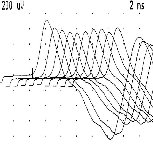
Electrodiagnostic studies may be supportive, and especially helpful in uncertain cases. These include repetitive stimulation studies. Essentially a repetitive electrical nerve stimulation is applied to a single muscle, and the amplitude of the compound muscle action potential measured.
In myasthenia gravis there will be a decrement of the amplitude on repeated stimulation, as a reflection of the progressively impaired conduction at the neuromuscular junction, and consequent fatigue, see figure 1.
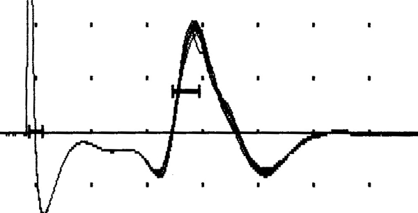
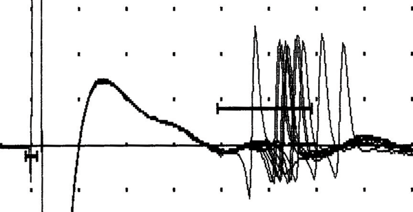
A single fibre EMG study may also be performed, and jitter or instability of the neuromuscular junction transmission should be detected, see figure 2. Single fibre EMG is reported to have a sensitivity of around 94% and specificity of around 79% for ocular myasthenia presenting with ptosis.3 For myasthenia gravis in general a sensitivity of 98% and specificity of 70% has been reported.4
An ice pack test may be used in a patient with ptosis. The ice pack is applied to the upper eyelid for 2-5 minutes, and an improvement in ptosis by 2 mm or more is considered a positive test. The ice pack test has a sensitivity of 86% and specificity of 79%.3
An edrophonium test is occasionally used where there is doubt. Edrophonium, a very short-acting acetylcholinesterase inhibitor, is administered intravenously and an improvement in signs is a positive result.
The main clinical presentations are ocular myasthenia (the presence of any ocular muscle weakness, the patient may have weakness of eye closure, but no other weakness), and generalised myasthenia gravis. Most patients with ocular myasthenia develop generalised myasthenia within three years.
Patients with MuSK antibodies may have a different clinical presentation that usually affects young women, with prominent bulbar (speech and swallowing) and respiratory symptoms, higher risk of developing myasthenic crisis, a more challenging course and more resistance to standard treatments.
Having established a diagnosis of myasthenia gravis, an MRI or CT scan of the thymus is performed, looking for possible thymoma that may be associated with the condition.
If the patient has respiratory symptoms, pulmonary function tests such as forced vital capacity, arterial blood gas, and spirometry assessment may be important.
Differential diagnosis
Other rarer myasthenic conditions to distinguish include Lambert-Eaton myasthenic syndrome (where the antibody is to the presynaptic voltage-gated calcium channel, and often associated with small cell lung carcinoma), and congenital myasthenic syndromes (these are a result of mutations in genes for neuromuscular junction proteins, and do not have an autoimmune basis).
Organophosphate poisoning from insecticides destroys the acetylcholinesterase, and botulism prevents the presynaptic release of acetylcholine, at the neuromuscular junction, causing acute presentations of neuromuscular junction dysfunction.
A wider range of neurological and neuromuscular conditions may also present with fatigue and weakness but are usually more readily distinguishable.
Potential complications
The potential complications relate to muscle fatigue, especially respiratory insufficiency and swallowing difficulties. In a patient with progressive respiratory insufficiency urgent hospital admission may be needed.
A rapid clinical worsening that could lead to crisis in the short term, days to weeks, indicates an impending myasthenic crisis. Approximately 20% of patients with myasthenia gravis experience a crisis in their lifetime, typically within two years of diagnosis.
An impending myasthenic crisis will probably require hospital admission for assessment, observation of respiratory and bulbar function, and ready access to an intensive care unit (ICU). Myasthenic crisis requires urgent and direct admission to ICU. Patients may require intubation or noninvasive ventilation to avoid intubation.
Deterioration may be triggered by a respiratory or other infection. A number of drugs, including aminoglycosides such as gentamicin and beta-blockers may cause deterioration. Deterioration is usually a result of undertreatment but may also be caused by excessive use of acetylcholinesterase inhibitors (a cholinergic crisis), and it may be difficult to distinguish between these situations.
Fifty years ago mortality in myasthenia gravis crisis was estimated to be between 20 and 50%. Today this figure is < 5%, due to improved intensive care and respiratory management, new immunomodulatory therapies, and importantly improved anticipatory management and early admission.5
Management
Pharmacotherapy
There are national UK6,7 and international8,9,10 guidelines on the management of myasthenia gravis. Management will usually be initiated by the neurologist, but as a chronic condition the GP will be involved in the long-term management and monitoring, usually with the patient continuing with neurological follow-up. The GP may also be the first point of contact for an acute deterioration.
First-line treatment is with pyridostigmine, an acetylcholinesterase inhibitor that prevents the breakdown of the neuromuscular transmitter acetylcholine at the neuromuscular junction, facilitating neuromuscular transmission, see table 1.
Dosage depends on the severity of symptoms, but initially may be commenced at 30-60 mg four times daily. The 60 mg tablets can be split in two. The half-life is short, and the effect of a single tablet lasts only a few hours. The cholinergic side effects may include abdominal cramps and diarrhoea. Propantheline or mebeverine may help with these side effects.
The dose of pyridostigmine will need to be adjusted according to the individual patient’s response and this varies considerably between patients. The dose is initially titrated to the lowest effective dose. Patients may become adept at adjusting their medication according to response and activity.
Pyridostigmine is a symptomatic treatment and does not treat the underlying cause of the autoimmune condition (usually antibodies to the acetylcholine receptor).
The next step in management would be with an immunosuppressant, usually prednisolone. In order to minimise the side effects of long-term steroids, a steroid-sparing agent such as azathioprine may be introduced.11
The activity of the enzyme thiopurine S-methyltransferase (TPMT) may be reduced in some patients. TPMT is involved in the metabolism of azathioprine, and patients with a reduced activity level are at higher risk of severe myelosuppression by the azathioprine. TPMT activity level is measured as a blood test before commencing azathioprine, as a lower dose may be needed.
Other immunosuppressants that may be considered include cyclosporine, mycophenolate, and methotrexate. Methotrexate is teratogenic. Women and men (there may be a possible trace in semen) are advised to use appropriate contraception during treatment and up to 6 months after cessation of treatment.
Medication to prevent steroid-induced osteoporosis is an important consideration, usually with reference to local guidelines. Azathioprine requires regular long-term monitoring of full blood count (in particular white cell count) and increasingly there
will be local guidelines on shared care for monitoring and prescribing. Prednisolone may induce or exacerbate diabetes. It is important to be aware that commencement of steroid threrapy may cause an initial deterioration in the myasthenia symptoms.
The majority of patients may be managed on this regimen, but some will require additional treatment, including intravenous immunoglobulin or plasma exchange for acute deterioration. Newer developments include more aggressive immunosuppression with drugs such as rituximab, and this would be directed by a specialist in myasthenia gravis.12
Other treatments
In a rare disease such as myasthenia gravis it is difficult to perform randomised clinical trials. It may also be difficult to generalise the results of these trials to individual patients. There is clinical trial evidence to indicate that thymectomy in non-thymomatous myasthenia gravis may be helpful in long-term management of selected patients with myasthenia gravis.13
The surgery may be performed using a thoracotomy or an endoscopic approach. Thymectomy is indicated in patients with myasthenia gravis and thymoma, this is primarily to remove the tumour and may not improve the myasthenia gravis.10
In patients with persistent disability related to muscle weakness, physiotherapy and other rehabilitation approaches are indicated. It is usually necessary to continue immunosuppression for many years, but with attempts at reduction where possible. There may be remission, and recurrence of myasthenia gravis.
Pregnancy
Pregnancy should be planned so that myasthenic treatment can be optimised and risk to the fetus minimised.6
The majority of women remain stable during pregnancy, and any worsening is more likely in the first few months after delivery. The aim is for spontaneous vaginal delivery, but it is important that the baby has rapid access to neonatal intensive care if needed.
In neonatal myasthenia, the newborn baby receives the acetylcholine receptor antibodies from a mother with myasthenia gravis. The neonatal symptoms may take several days to develop and require careful observation.
Conclusion
Myasthenia gravis is a rare autoimmune disorder affecting neuromuscular junction function. In its most frequent presentation it should be considered, and provisionally diagnosed, by the GP on the basis of a clinical history of fatiguing muscle weakness and basic clinical examination. Patients should be referred to a neurologist to confirm diagnosis and initiate management.
Myasthenia gravis is a chronic condition, and over the years monitoring and adjustment of treatment will be required. Monitoring of the potential rare complications of immunosuppressant treatment is vital.
Most important is the recognition of impending myasthenic crisis (for which there should be a low threshold for hospital admission), and for myasthenic crisis for which urgent admission to ICU is essential, to minimise preventable deaths from the condition.
REFERENCES
1 Vincent A. Antibodies and receptors: from neuromuscular junction to central nervous system. Neuroscience 2020;439:48-61
2 Robertson NP, Deans J, Compston DAS. Myasthenia gravis: a population based epidemiological study in Cambridgeshire, England. J Neurol Neurosurg Psychiatry 1998;65:492-96
3 Ciannoccaro MP, Paolucci M, Zenesini C et al. Comparison of ice-pack test and single fiber EMG diagnostic accuracy in patients referred for myasthenic ptosis. Neurology 2020. (Online ahead of print)
4 Padua L, Caliandro P, Di Iasi G et al. Reliability of SFEMG in diagnosing myasthenia gravis: sensitivity and specific calculated on 100 prospective cases. Clin Neurophysiol 2014;125:1270-73
5 Alshekhlee A, Miles JD, Katirji B et al. Incidence and mortality rates of myasthenia gravis and myasthenic crisis in US hospitals. Neurology 2009;72:1548-54
6 Norwood F, Dhanjal M, Hill M et al. Myasthenia in pregnancy: best practice guidelines from a UK multispecialty working group. J Neurol Neurosurg Psychiatry 2014;85:538-43
7 Sussman J, Farrugia ME, Maddison P et al. Myasthenia gravis: Association of British Neurologists’ management guidelines. Pract Neurol 2015;15:199-206
8 Elsais A, Argove Z, Evoli A et al. EFNS/ENS guidelines for the treatment of ocular myasthenia. Eur J Neurol 2014;21:687-93
9 Sanders DB, Wolfe GI, Benatar M et al. International consensus guidance for management of myasthenia gravis: Executive summary. Neurology 2016;87:419-25
10 Gronseth GS. Barohn R, Narayanaswami P. Practice advisory: thymectomy for myasthenia graivs (practice parameter update). Report of the Guideline Development, Dissemination, and Implementation Subcommittee of the American Academy of Neurology. Neurology 2020;94:705-09
11 Palace J, Newsom Davis J, Lecky B. A randomized double-blind trial of prednisolone alone or with azathioprine in myasthenia gravis. Myasthenia Gravis Study Group. Neurology 1998;50:1778-83
12 Dalakas MC. Progress in the therapy of myasthenia gravis: getting closer to effective targeted immunotherapies. Curr Opin Neurol 2020 [Online ahead of print]
13 Wolfe GJ, Kaminski HJ, Aban IB et al. Long-term effect of thymectomy plus prednisone alone in patients with non-thymomatous myasthenia gravis: 2-year extension of the MGTX randomised trial. Lancet Neurol 2019;18:259-68
 Download this article in PDF format