SEARCH

Lau MS, Hopper AD, Sanders DS. Improving the detection of coeliac disease. Practitioner 2016;260(1795):13-17
Improving the detection of coeliac disease
01 Aug 2016
AUTHORS
Dr Michelle S Lau MBChB MRCP, Clinical Research Fellow in Gastroenterology
Dr Andrew D Hopper MD FRCP, Gastroenterology Consultant
Professor David S Sanders FRCP MD FACG, Professor of Gastroenterology, Academic Unit of Gastroenterology, Royal Hallamshire Hospital & University of Sheffield, Sheffield, UK
Article
Abstract
The common presentation of coeliac disease has shifted from the historically classical symptoms of malabsorption in childhood to non-classical symptoms in adulthood such as irritable bowel syndrome-type symptoms, anaemia, chronic fatigue, change in bowel habit, abdominal pain and osteoporosis. A combination of coeliac serology and duodenal biopsy is required to diagnose coeliac disease in adults. Testing for IgA-tissue transglutaminase antibodies should be carried out as a first-line screening test. Advise patients to eat a gluten-containing diet for six weeks before their investigations to ensure the serological and histological results are not affected. A confirmatory duodenal biopsy is mandatory to ensure that patients are correctly diagnosed with coeliac disease. A lifelong strict gluten-free diet is the only effective treatment currently available. All patients should be referred to a specialist dietitian for guidance and support. Annual follow-up can begin when the disease is stable and patients are managing well on their diet.
It has been increasingly recognised over the past few years that coeliac disease presents in a much wider range of manifestations than previously thought. The common presentation has shifted from the historically classical symptoms of malabsorption in childhood to non-classical symptoms in adulthood.
Classical symptoms include chronic diarrhoea, weight loss and failure to thrive,2 which are relatively rare. The more common non-classical symptoms include irritable bowel syndrome-type symptoms,anaemia, chronic fatigue, change in bowel habit, abdominal pain and osteoporosis.3,4
Population screening studies have shown that the prevalence of coeliac disease is 1 in 100,3,4 and yet 75% of patients remain undiagnosed.5 A recent study demonstrated significantly impaired quality of life in patients living with undiagnosed coeliac disease compared with those who have been diagnosed and treated (EQ-5D 0.56 versus 0.84).6
Table 1 lists the symptoms or risk factors which should trigger screening for coeliac disease according to the recently updated NICE guidelines.1
Investigation
A combination of coeliac serology and duodenal biopsy is required for the diagnosis of coeliac disease in adults.
The current British Society of Gastroenterology7 and NICE1 guidelines recommend screening high-risk adults by coeliac serology. Testing for IgA-tissue transglutaminase antibodies (IgA-TTG) should be performed as a first-line screening test, as it has a high sensitivity and negative predictive value.
A study of 2,000 patients with a prevalence of coeliac disease of 3.4% compared the sensitivities of IgA-TTG alone and a two-step approach testing for IgA-TTG and endomysial antibodies (EMA). IgA-TTG alone was found to be a sensitive marker for coeliac disease compared with IgA-TTG plus EMA (sensitivity 90.9% vs 85.7%, and negative predictive value 99.6% vs 99.7%).8 Therefore, IgA-TTG is the recommended first-line test. However, if IgA-TTG is weakly positive, EMA testing should be carried out.7 This has a high specificity of 95%.9,10 It should be noted that EMA testing utilises the substrates of monkey oesophagus or human umbilicus, which have limited availability. It is also expensive, labour intensive and interpretation of the results is subjective.
Patients with IgA deficiency do not produce IgA-TTG or IgA-EMA antibodies, and could therefore give a false-negative result. IgA deficiency is ten times more prevalent in coeliac disease compared with the general population (2.6% vs 0.14-0.2%).11
Therefore it is essential to test for immunoglobulin levels in conjunction with serology routinely. For those with IgA deficiency, IgG-TTG, IgG-EMA and IgG-deamidated gliadin peptide (IgG-DGP) can be tested instead.
A recent meta-analysis found that IgG-DGP had a pooled sensitivity ranging from 80.1% to 98.6% compared with TTG ≥ 90%.12
In recent years, several point of care tests have become commercially available in pharmacies and online for patients to purchase.13,14 However, there is relatively limited data in the literature on their performance compared with conventional serology. Therefore, the results should be confirmed with TTG and EMA.
It is important to advise patients to eat a gluten-containing diet (10 g of gluten a day, equivalent to four slices of bread) for six weeks before their investigations to ensure the serological and histological results are not affected.
Diagnosis
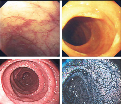
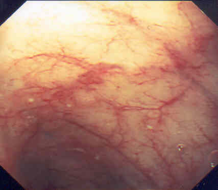
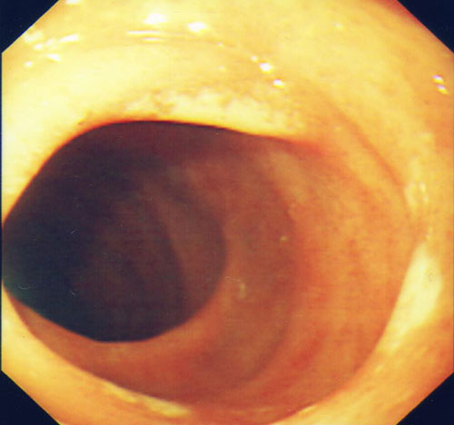
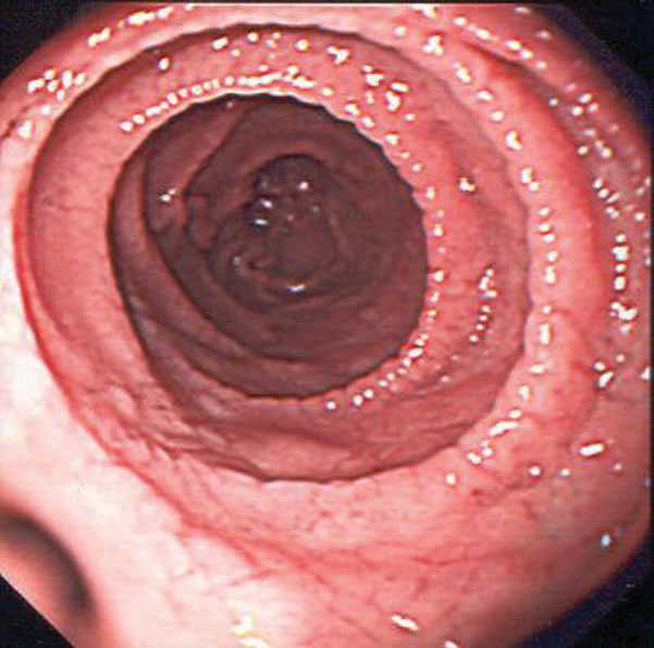
Duodenal biopsies showing increased intraepithelial lymphocytes, crypt hyperplasia and villous atrophy (Marsh 3a-3c) with positive coeliac serology confirms the diagnosis of coeliac disease in adults.
Although studies have demonstrated excellent sensitivities for IgA-TTG and EMA, the sensitivities often drop when the tests are performed in the real world with a lower prevalence in the general population.
Therefore, a confirmatory duodenal biopsy is mandatory to ensure that patients are correctly diagnosed with coeliac disease before being subjected to a lifelong gluten-free diet. Additionally, it avoids any diagnostic uncertainty in cases where patients do not respond to a gluten-free diet, and provides a baseline reference for histological response to a gluten-free diet.
It must be noted that clinical response to a gluten-free diet does not confer a diagnosis of coeliac disease. This can be seen in patients with non-coeliac gluten sensitivity, which is characterised by symptom improvement on gluten avoidance without coeliac disease (negative coeliac serology and absence of villous atrophy).15
On the other hand, the recent European Society for Paediatric Gastroenterology Hepatology and Nutrition guidelines recommend that paediatric patients no longer require biopsies to confirm the diagnosis of coeliac disease if they have TTG levels ten times over the upper limit of normal, positive EMA and the presence of the HLA-DQ2 or HLA-DQ8 genotype.16
This avoids the need for a gastroscopy which often requires general anaesthesia in the paediatric population. This is an appropriate approach in carefully selected paediatric cases in the hands of specialists but should not be extended to adult practice. With the poor specificity of TTG and variation in assays across the UK, the potential risk is of adults being committed to a gluten-free diet who do not have coeliac disease.
Referral
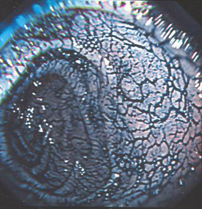
All adult patients with positive coeliac serology should be referred to gastroenterology for a gastroscopy, see figure 1, to confirm or exclude coeliac disease. Children with positive coeliac serology should be referred to paediatric gastroenterology for further investigations.
Any patients with negative serology should be referred to gastroenterology if coeliac disease is still clinically suspected, as seronegative coeliac disease occurs in 7% of patients.17
‘Advise patients to eat a gluten containing diet for six weeks before their investigations to ensure the serological and histological results are not affected’
IgA deficiency needs to be ruled out as a cause for negative IgA-TTG or EMA. Immunosuppressants and steroids can also cause seronegativity. A highly accurate clinical decision algorithm developed by Hopper and colleagues demonstrated that as long as the patient did not have high-risk symptoms of anaemia, weight loss or diarrhoea, coeliac disease could be safely ruled out by negative serology.
A negative HLA DQ2 or DQ8 genotype could be useful in this situation to exclude coeliac disease, as these genotypes are expressed in virtually all patients with coeliac disease. However, the presence of these genotypes does not confirm the diagnosis of coeliac disease, because they are also present in 25-40% of the general population.18
If HLA DQ2 or DQ8 is present in seronegative patients with a clinical suspicion of coeliac disease duodenal biopsy should be performed.19
Management
A lifelong strict gluten-free diet is the only effective treatment currently available. All patients should be referred to a specialist dietitian for thorough guidance and support.
A gluten-free diet can pose challenges in terms of adherence for some individuals. Moreover, difficulties may be encountered when eating out or eating separately in the same household.
A systematic review showed that up to 41% of patients were found not to be strictly adhering to a gluten-free diet.20 Patients should be encouraged to join coeliac disease advocacy groups (see Useful information box), as evidence shows that this is strongly associated with strict dietary adherence.20
The goal of treatment is to relieve symptoms, achieve mucosal healing to prevent complications, and improve quality of life. Adherence assessment includes a combination of dietetic evaluation, a validated adherence questionnaire, symptom assessment, serological testing and repeat duodenal biopsy. Apart from duodenal histology being the gold standard for assessing for mucosal recovery, none of the above methods alone are reliable markers of mucosal healing, with sensitivities ranging from 38 to 61%.21,22
Internationally, a repeat biopsy to assess for histological remission is undertaken, but this is not routine practice in the UK.7 A recent study of 7,648 patients with coeliac disease over an 11 year follow-up period showed that patients with persistent villous atrophy had an increased incidence of lymphoproliferative malignancies compared with those without (HR 3.61). However, there was no difference in the incidence of lymphoproliferative malignancies between the re-biopsy and no biopsy groups, thereby questioning the role of a routine follow- up biopsy in all patients.23
In the UK, a follow-up biopsy should be considered in patients who do not respond to a gluten-free diet, for instance those with persistent symptoms or persistently raised serological titres despite strict dietary adherence. This would help identify patients with persistent villous atrophy who may require immunosuppressants in cases of refractory coeliac disease, and early detection of enteropathy associated T-cell lymphoma which carries a poor prognosis.
Annual follow-up can begin when the disease is stable and patients are managing well on their diet. Small bowel absorption should be assessed by measuring full blood count, ferritin, folate and B12 levels. There is an increased risk of osteoporosis and fractures, which improve with gluten exclusion. Calcium, vitamin D and alkaline phosphatase should be monitored and supplemented accordingly. Those with additional risk factors for osteoporosis or age over 55 should have a baseline bone density scan one year after starting a gluten-free diet. Other co-existing autoimmune diseases should be screened for, such as autoimmune thyroid disease and type 1 diabetes.
Liver function tests should also be performed as coeliac disease can be associated with deranged liver enzymes. A recent study showed that liver function was deranged in 41% of patients with coeliac disease at diagnosis, which normalised in 78% of patients 1.5 years after gluten exclusion. Those without any improvement in liver function should be tested for any
co-existing liver disease.24 Functional hyposplenism is seen in patients with coeliac disease, which may lead to impaired immunity to encapsulated bacteria.
Data showed a modest increase in pneumococcal sepsis in patients with coeliac disease compared with the general population (HR 2.6),25 therefore pneumococcal vaccination is recommended every five years after the initial vaccination.
Follow-up
Patients with no symptoms and a normal duodenal biopsy are at low risk of complications and can be safely discharged to the community.26
Serology is frequently used as a surrogate marker of histological recovery although the relationship is not linear and normal serology does not rule out persistent villous atrophy.26,27 Those with either symptoms or villous atrophy or both should be followed up in secondary care to assess for dietary adherence and/or to investigate the cause of persistent villous atrophy.
Non-responsive coeliac disease describes patients who do not respond after 6-12 months on a gluten-free diet or relapse with symptoms despite gluten avoidance. The most common reason for unresolved symptoms is persistent gluten exposure, therefore the patient should undergo a thorough dietetic assessment to identify any gluten exposure or cross contamination.20 Unresolved symptoms can also be caused by other gastrointestinal conditions which are associated with coeliac disease, such as small bowel bacterial overgrowth, pancreatic insufficiency and microscopic colitis.28 The validity of the initial diagnosis of coeliac disease should be reviewed, and other associated conditions as mentioned before should be ruled out.
Refractory coeliac disease occurs in a very small minority of patients with persistent symptoms. It is a rare condition which is defined by persistent villous atrophy despite strict adherence for 12 months to a gluten-free diet, after eliminating other causes of non-responsive coeliac disease.
Patients with refractory coeliac disease commonly suffer from weight loss, diarrhoea, abdominal pain, nutritional deficiencies and anaemia.29 They are at risk of developing enteropathy associated T-cell lymphoma, and require urgent evaluation at a tertiary centre.
A flow chart for the management of non-responsive coeliac disease is shown in figure 2.
REFERENCES
1 National Institute for Health and Care Excellence. NG20. Coeliac disease: recognition, assessment and management. NICE. London. 2015
2 Campbell CB, Roberts RK, Cowen AE. The changing clinical presentation of coeliac disease in adults. Med J Aust 1977;1(4):89-93
3 West J, Logan RF, Hill PG et al. Seroprevalence, correlates, and characteristics of undetected coeliac disease in England. Gut 2003;52(7):960-5
4 Fasano A, Berti I, Gerarduzzi T et al. Prevalence of celiac disease in at-risk and not-at-risk groups in the United States: a large multicenter study. Arch Int Med 2003;163(3):286
5 West J, Fleming KM, Tata LJ et al. Incidence and prevalence of celiac disease and dermatitis herpetiformis in the UK over two decades: population-based study. Am J Gastroenterol 2014;109(5):757-68
6 Gray AM, Papanicolas IN. Impact of symptoms on quality of life before and after diagnosis of coeliac disease: results from a UK population survey. BMC Health Services Res 2010;10:105
7 Ludvigsson JF, Bai JC, Biagi F et al. Diagnosis and management of adult coeliac disease: guidelines from the British Society of Gastroenterology. Gut 2014;63(8):1210-28
8 Hopper AD, Hadjivassiliou M, Hurlstone DP et al. What is the role of serologic testing in celiac disease? A prospective, biopsy-confirmed study with economic analysis. Clin Gastroenterol Hepatol 2008;6(3):314-20
9 Leffler DA, Schuppan D. Update on serologic testing in celiac disease. Am J Gastroenterol 2010;105(12):2520-24
10 Lewis NR, Scott BB. Systematic review: the use of serology to exclude or diagnose coeliac disease (a comparison of the endomysial and tissue transglutaminase antibody tests). Aliment Pharmacol Therap 2006;24(1):47-54
11 Cataldo F, Marino V, Ventura A et al. Prevalence and clinical features of selective immunoglobulin A deficiency in coeliac disease: an Italian multicentre study. Italian Society of Paediatric Gastroenterology and Hepatology (SIGEP) and "Club del Tenue" Working Groups on Coeliac Disease. Gut 1998;42(3):362-65
12 Giersiepen K, Lelgemann M, Stuhldreher N et al. Accuracy of diagnostic antibody tests for coeliac disease in children: summary of an evidence report. J Pediatr Gastroenterol Nutr 2012;54(2):229-41
13 Mooney PD, Kurien M, Sanders DS. Simtomax, a novel point of care test for coeliac disease. Expert Opin Med Diag 2013;7(6):645-51
14 Korponay-Szabo IR, Szabados K, Pusztai J et al. Population screening for coeliac disease in primary care by district nurses using a rapid antibody test: diagnostic accuracy and feasibility study. BMJ 2007;335(7632):1244-47
15 Biesiekierski JR, Newnham ED, Irving PM et al. Gluten causes gastrointestinal symptoms in subjects without celiac disease: a double-blind randomized placebo-controlled trial. Am J Gastroenterol 2011;106(3):508-14
16 Husby S, Koletzko S, Korponay-Szabó IR et al. European Society for Pediatric Gastroenterology, Hepatology, and Nutrition guidelines for the diagnosis of coeliac disease. JPGN 2012;54:136-60
17 Lewis NR, Scott BB. Meta-analysis: deamidated gliadin peptide antibody and tissue transglutaminase antibody compared as screening tests for coeliac disease. Aliment Pharmacol Ther 2010;31(1):73-81
18 Hadithi M, Von Blomberg BME, Crusius JBA et al. Accuracy of serologic tests and HLA-DQ typing for diagnosing celiac disease. Ann Int Med 2007;147(5):294
19 Hopper AD, Cross SS, Hurlstone DP et al. Pre-endoscopy serological testing for coeliac disease: evaluation of a clinical decision tool. BMJ 2007;334(7596):729
20 Hall NJ, Rubin G, Charnock A. Systematic review: adherence to a gluten-free diet in adult patients with coeliac disease. Aliment Pharmacol Therap 2009;30(4):315-30
21 Rubio-Tapia A, Murray JA. Classification and management of refractory coeliac disease. Gut 2010;59(4):547-57
22 Biagi F, Andrealli A, Bianchi PI et al. A gluten-free diet score to evaluate dietary compliance in patients with coeliac disease. Br J Nutr 2009;102(6):882-87
23 Lebwohl B, Granath F, Ekbom A et al. Mucosal healing and risk for lymphoproliferative malignancy in celiac disease: a population-based cohort study. Ann Int Med 2013;159(3):169
24 Natalia EC, Rohini RV, Thimmaiah GT et al. Prevalence of abnormal liver function tests in celiac disease and the effect of a gluten- free diet in the US population. Am J Gastroenterol 2015;110(8):1216
25 Ludvigsson JF, Olen O, Bell M et al. Coeliac disease and risk of sepsis. Gut 2008;57(8):1074-80
26 Sharkey LM, Corbett G, Currie E et al. Optimising delivery of care in coeliac disease - comparison of the benefits of repeat biopsy and serological follow-up. Aliment Pharmacol Ther 2013;38(10):1278-91
27 Kaukinen K, Sulkanen S, Maki M, Collin P. IgA-class transglutaminase antibodies in evaluating the efficacy of gluten-free diet in coeliac disease. Eur J Gastroenterol Hepatol 2002;14(3):311-15
28 Rubio-Tapia A, Hill ID, Kelly CP et al. American College of Gastroenterology. ACG clinical guidelines: diagnosis and management of celiac disease. Am J Gastroenterol 2013;108(5):656-76
29 Al-toma A, Verbeek WH, Mulder CJ. The management of complicated celiac disease. Dig Dis 2007;25(3):230-36
ARTICLE IN PDF
 Download this article in PDF format
CLICK ON IMAGES
PRESET SEARCHES
PubMed
NICE evidence search
EXTERNAL WEBLINKS
Coeliac UK