SEARCH

The Practitioner 2011 255 (1742):15-19
Upper GI bleeding requires prompt investigation
08 Aug 2011
AUTHORS
Dr Andrew D Hopper MD MRCP
Gastroenterology Consultant, Department of Gastroenterology, Royal Hallamshire Hospital, Sheffield
Professor David S Sanders MD FRCP FACG, Gastroenterology Consultant, Department of Gastroenterology, Royal Hallamshire Hospital and University of Sheffield
Article
Upper GI bleeding is a common medical emergency with an incidence in the UK of 103 cases per 100,000 adults per year and is much more common in the elderly.1
A national audit by the British Society of Gastroenterology (BSG) on the management of upper GI bleeding in the UK, conducted between 1993 and 1994, showed 30-day mortality to be 14%.2 Based on this audit several recommendations were made, including urgent endoscopy in high-risk patients and admission or early referral of patients with upper GI bleeding to gastroenterology teams. However, despite improvements in endoscopy, recognition of provoking factors and pharmacological interventions available when the audit was repeated in 2007 the mortality rate still remained significant at 10%.3
Presentation
The most common presenting signs of acute upper GI bleeding are haematemesis, either bright red or ‘coffee ground', and melaena. About 30% of patients with bleeding ulcers present with haematemesis, 20% with melaena, and 50% with both. Haematochezia usually suggests a lower GI source of bleeding, however, up to 5% of patients with bleeding ulcers have haematochezia and it indicates heavy bleeding into the upper GI tract.4 An upper GI bleeding source should be considered when haematochezia presents with signs and symptoms of haemodynamic compromise such as syncope, postural hypotension, tachycardia, and shock should therefore direct one's attention to an upper GI source of bleeding.
More insidious presentations would include nausea, vomiting, epigastric pain, vasovagal phenomena, and syncope, however systemic symptoms such as these would be rare without a history of melaena.
Common causes
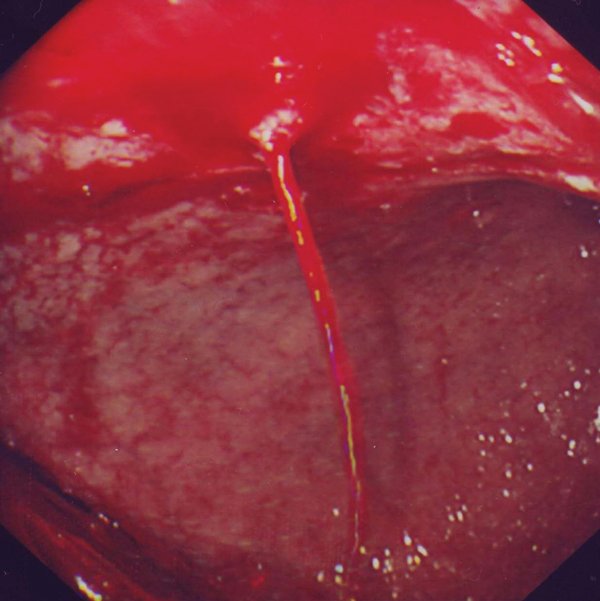
Peptic ulcer disease, both gastric and duodenal, accounts for the majority of admissions for upper GI bleeding, see table 1, above. Significant bleeding can result from involvement of large vessels within the base of the ulcer see figure 1, p15.3 The main risk factors for developing peptic ulcers are a history of NSAID use 5 or the presence of Helicobacter pylori infection.6 Warfarin and clopidogrel use can potentiate bleeding, however clopidogrel can also induce bleeding independently in patients with previous peptic ulcer disease.7
Bleeding from oesophageal varices usually results in large volumes of bright red haematemesis, if a previous history of chronic liver disease (with or without varices) is known or signs of chronic liver disease are present then an upper GI bleed should be treated as a serious emergency regardless of cardiovascular stability.
Other causes of bleeding are listed in table 1, above, these include a mucosal (Mallory-Weiss) tear of the gastro-oesophageal junction secondary to vomiting, and multiple types of vascular abnormalities. Apart from variceal disease all other causes can vary in severity and with assessment and risk stratification appropriate initial management, early endoscopy or early discharge can be planned.
Assessment
Clinical risk factors for mortality in upper GI bleeding are: age; comorbidity; tachycardia and a low systolic blood pressure and should be assessed at presentation. The Rockall scoring system incorporates these clinical variables and can be used to predict mortality risk from an ‘initial' pre-endoscopy score (see tables 2 and 3, opposite). This system is used widely in the UK. It was developed from a large prospective audit and has been validated numerous times.8 If the endoscopy result is known then a ‘full' score can also be used. As well as mortality the risk of re-bleeding can also be predicted which is usually used following endoscopy to aid more intensive therapy/observation decisions if high, see table 3, opposite. The benefits of this scoring system are not only that mortality can be predicted but also if initial scores are zero, patients suitable for early discharge with outpatient endoscopy can be identified, or admission avoided altogether.
When a delay in presentation of a haematemesis or melaena (over a number of days) occurs in a low-risk patient then a discussion with the local gastroenterology team to arrange urgent (within 24-48 hours) outpatient endoscopy can prevent hospital admission. In these cases access to up-to-date haemoglobin and urea results is especially useful,9 however, nearly all patients with upper GI bleeding symptoms should be referred to secondary care for further assessment, endoscopy and intervention as required.
Referral
Given the high mortality rate associated with upper GI bleeding nearly all patients with symptoms described above should be referred to secondary care for emergency admission and endoscopic assessment. This should also be the default position in borderline cases. Only young patients (
Therapeutic endoscopy after upper GI bleeding has been clearly shown to reduce re-bleeding and the need for surgery and consequently there has been a push to provide timely endoscopy services for upper GI bleeding patients nationally.10 A previous audit carried out in 1993 by the BSG found delays in undertaking endoscopy, leading the BSG guidelines to recommend that all medium- to high-risk patients with acute upper GI bleeding undergo endoscopy within 24 hours.1 In a recent update of the audit 52% of hospitals provided a consultant-led out of hours endoscopy service. The risk adjusted mortality ratio was higher (1.51 versus 1.21) but not significant (p=0.1) in hospitals without such rotas. Individual reports have shown that out of hours endoscopy with a dedicated bleed unit may be able to reduce mortality significantly further.11
Early endoscopy in upper GI bleeding:
- allows early diagnosis
- provides the opportunity for endoscopic haemostasis
- enables complete risk stratification of non-variceal bleeding
- allows early discharge of patients with low-risk findings
- has resulted in a reduction in transfusion requirements and length of stay for all risk groups without cost shifting to the outpatient department.12,13
However, no study has been able to demonstrate that early endoscopy leads to a reduction in mortality, this is probably because the major determinant of survival is comorbidity rather than control of haemostasis.8,14 Nevertheless the BSG guidelines along with others emphasise the importance of endoscopy within 24 hours of presentation particularly for high-risk patients.15
Patients failing to be controlled with endoscopy can have either interventional radiology with vessel embolisation or surgery. In the recent BSG audit this occurred in 1.7% and 2.2% of patients with upper GI bleeding respectively. The mortality rate in the patients who underwent surgery with upper GI bleeding uncontrolled by endoscopy was 31%.
Pharmacological options
Approximately one third of patients who present with a bleeding ulcer will develop recurrent bleeding within two years if left untreated after ulcer healing.16 All patients should be discharged on an oral proton pump inhibitor (PPI), with dosage and duration dependent on the underlying aetiology, to decrease the risk of re-bleeding. All patients with a gastric ulcer should be considered for a repeat endoscopy at six weeks to check healing given the possible presentation of an early gastric cancer as a gastric ulcer. Subsequent secondary prevention for upper GI bleeding at discharge aims to prevent recurrence and management varies depending on the aetiology.
Helicobacter pylori eradication
The H. pylori infection rate in patients with bleeding peptic ulcers has been calculated as 80%.17 Patients with peptic ulcer disease from H. pylori who are not using NSAIDs should undergo successful eradication and a further three weeks of PPI therapy.18 In patients who have a history of NSAID or aspirin use continuation of PPI therapy after this period is discussed below.
Biopsy samples to test for the presence of H. pylori should be taken at initial endoscopy if possible before commencing PPI therapy. Eradication should be confirmed by breath test or biopsy to minimise the risk of re-bleeding from peptic ulcers. In the case of eradication failure, after compliance discussion, second-line treatment should be prescribed, see table 4, p18.
H. pylori testing to confirm successful eradication should only be undertaken after PPI and antibiotic therapy has been completed and discontinued since testing within two weeks of these treatments (especially urease biopsy methods) may result in false-negative findings.19 Repeat testing can of course be conveniently timed with the repeat endoscopy in cases of gastric ulcer disease.
There is no evidence to support empirical eradication of H. pylori in patients with bleeding peptic ulcers and this should only occur if exceptional circumstances prevent testing.
NSAIDs
Patients having experienced a bout of upper GI bleeding while on an NSAID should have their treatment discontinued or replaced by an NSAID with PPI cover.
Clinical factors reported to increase the risk of developing NSAID-associated upper GI complications include:
- a history of ulcer or GI bleeding
- increasing age
- concomitant anticoagulation
- concomitant corticosteroid therapy
- high-dose NSAID use.
The risk associated with the NSAID persists for approximately two months after the treatment is stopped, therefore PPI therapy of adequate duration should be considered.20
PPI and eradication of H. pylori infection are options to use in combination with NSAIDs in situations where it is necessary to continue the NSAIDs. Recurrent bleeding after six months among aspirin users has been shown in 1.9% of patients after H. pylori eradication therapy and 0.9% on long-term omeprazole (20 mg od). The aspirin dose used in the study ranged from 80 to 325 mg/day but was mainly low dose (mean 101 mg/day, n=250). Among naproxen users, respective figures are 18.8% after eradication therapy and 4.4% on long-term omeprazole (20 mg). Therefore PPIs should be used in preventing recurrent bleeding in patients who are taking non-aspirin NSAIDs but the risk (4.4%) should still be noted.21
Aspirin and clopidogrel
Aspirin therapy is associated with a dose-related risk. Aspirin doubles the risk of upper GI bleeding at daily doses of 75 mg and quadruples it at doses of 300 mg.22 Enteric-coated formulations do not appear to reduce the risk.23
Patients requiring low-dose aspirin should resume taking aspirin as soon as the cardiovascular risks outweigh the risk of re-bleeding. This could be as early as five days after the onset of bleeding as the delay to a thrombotic event is generally reported as between seven and ten days.24 25 A patient who has had an episode of non-variceal upper GI bleeding while taking aspirin and who still requires cardioprotection should take PPI prophylaxis with aspirin rather than clopidogrel alone.
The safety of clopidogrel compared with a combination of aspirin and esomeprazole has been evaluated in two randomised controlled trials involving patients with previous aspirin-induced peptic ulcer bleeding.26,27 In one trial the cumulative incidence of recurrent bleeding during the 12-month period was 8.6% in the clopidogrel group and 0.7% in those taking aspirin 80 mg plus esomeprazole 20 mg twice daily.27
In patients who have bled on clopidogrel and are thus at a very high risk for re-bleeding, the benefits of PPI secondary prophylaxis can be used. Recent studies have shown that omeprazole decreases the antiplatelet effect of clopidogrel, possibly by inhibiting the CYP2C19 enzyme.28 However, concomitant use of pantoprazole, lansoprazole, and esomeprazole did not have this effect, suggesting that the interaction between PPI and clopidogrel is not a class effect.29,30
COX-2 inhibitors
The safety of a COX-2 inhibitor compared with a combination of a non-selective NSAID and a PPI has been evaluated with similar rates of re-bleeding ulcers found at six months.31 One randomised controlled trial demonstrated a significantly lower rate of recurrent upper GI bleeding with a COX-2 inhibitor plus a PPI (0%) compared with a COX-2 inhibitor alone (8.9%) over one year.32 However two meta-analyses have demonstrated an excess risk of serious cardiovascular events associated with COX-2 inhibitors compared with placebo.33,34 Recent guidelines endorsed by the BSG suggest COX-2 inhibitors are not recommended in patients with cardiovascular risk. If cardiovascular risk is a concern, naproxen with a PPI is recommended when alternative analgesic therapies fail.15
Oesophageal varices
The mainstay of pharmacological treatment of portal hypertension is nonselective beta-blockade with propanolol. This improves mortality and re-bleeding risk in oesophageal varices.35 Nitrates can be added or substituted if required.36,37 Most centres should offer repeat endoscopy with further banding to help eradicate the varices as well. Patients with varices should also be on PPI therapy to help prevent oesophagitis which can lead to re-bleeding. The one-year mortality of variceal bleeding is up to 80% mostly due to the underlying liver disease so most management is directed towards maintaining liver function.38,39
Conclusion
Upper GI bleeding is a common medical emergency with significant mortality and remains a considerable challenge. Rapid appropriate assessment and referral for early endoscopy can prevent morbidity and possibly reduce mortality. The use of reliable scoring systems has enabled early discharge and an evidence-based approach to subsequent secondary prevention can avoid recurrence.
REFERENCES
1 Rockall TA, Logan RF, Devlin HB et al. Incidence of and mortality from acute upper gastrointestinal haemorrhage in the United Kingdom. Steering Committee and members of the National Audit of Acute Upper Gastrointestinal Haemorrhage. BMJ 1995;311:222-6
2 Rockall TA, Logan RF, Devlin HB et al. Influencing the practice and outcome in acute upper gastrointestinal haemorrhage. Steering Committee of the National Audit of Acute Upper Gastrointestinal Haemorrhage. Gut 1997;41:606-11
3 Hearnshaw SA, Logan RF, Lowe D et al. Acute upper gastrointestinal bleeding in the UK: patient characteristics, diagnoses and outcomes in the 2007 UK audit. Gut Published online first 13 April 2011
4 Wara P, Stodkilde H. Bleeding pattern before admission as guideline for emergency endoscopy. Scand J Gastroenterol 1985;20:72-8
5 Hawkey CJ. Non-steroidal anti-inflammatory drug gastropathy: causes and treatment. Scand J Gastroenterol Supplement 1996;220:124-7
6 Rosenstock S, Jorgensen T, Bonnevie O et al. Risk factors for peptic ulcer disease: a population based prospective cohort study comprising 2416 Danish adults. Gut 2003;52:186-93
7 Ng FH, Wong SY, Chang CM et al. High incidence of clopidogrel-associated gastrointestinal bleeding in patients with previous peptic ulcer disease. Aliment Pharmacol Therap 2003;18:443-9
8 Rockall TA, Logan RF, Devlin HB et al. Risk assessment after acute upper gastrointestinal haemorrhage. Gut 1996;38:316-21
9 Blatchford O, Murray WR, Blatchford M. A risk score to predict need for treatment for upper-gastrointestinal haemorrhage. Lancet 2000;356:1318-21
10 Calvet X, Vergara M, Brullet E et al. Addition of a second endoscopic treatment following epinephrine injection improves outcome in high-risk bleeding ulcers. Gastroenterology 2004;126:441-50
11 Sanders DS, Perry MJ, Jones SG et al. Effectiveness of an upper-gastrointestinal haemorrhage unit: a prospective analysis of 900 consecutive cases using the Rockall score as a method of risk standardisation. Euro J Gastroenterol Hepatol 2004;16:487-94
12 Lee JG, Turnipseed S, Romano PS et al. Endoscopy-based triage significantly reduces hospitalization rates and costs of treating upper GI bleeding: a randomized controlled trial. Gastrointest Endosc 1999;50:755-61
13 Spiegel BM, Vakil NB, Ofman JJ. Endoscopy for acute nonvariceal upper gastrointestinal tract hemorrhage: is sooner better? A systematic review. Arch Intern Med 2001;161:1393-404
14 Blatchford O, Davidson LA, Murray WR et al. Acute upper gastrointestinal haemorrhage in west of Scotland: case ascertainment study. BMJ 1997;315:510-4
15 Palmer K, Nairn M. Management of acute gastrointestinal blood loss: summary of SIGN guidelines. BMJ 2008;337:a1832
16 Gisbert JP, Khorrami S, Carballo F et al. H. pylori eradication therapy vs. antisecretory non-eradication therapy (with or without long-term maintenance antisecretory therapy) for the prevention of recurrent bleeding from peptic ulcer. Cochrane Database Syst Rev 2004:CD004062
17 Gisbert JP, Pajares JM. Helicobacter pylori and bleeding peptic ulcer: what is the prevalence of the infection in patients with this complication? Scand J Gastroenterol 2003;38:2-9
18 Liu CC, Lee CL, Chan CC et al. Maintenance treatment is not necessary after Helicobacter pylori eradication and healing of bleeding peptic ulcer: a 5-year prospective, randomized, controlled study. Arch Intern Med 2003;163:2020-4
19 Guell M, Artigau E, Esteve V et al. Usefulness of a delayed test for the diagnosis of Helicobacter pylori infection in bleeding peptic ulcer. Aliment Pharmacol Therap 2006;23:53-9
20 Hernandez-Diaz S, Rodriguez LA. Association between nonsteroidal anti-inflammatory drugs and upper gastrointestinal tract bleeding/perforation: an overview of epidemiologic studies published in the 1990s. Arch Intern Med 2000;160:2093-9
21 Chan FK, Chung SC, Suen BY et al. Preventing recurrent upper gastrointestinal bleeding in patients with Helicobacter pylori infection who are taking low-dose aspirin or naproxen. N Engl J Med 2001;344:967-73
22 Weil J, Colin-Jones D, Langman M et al. Prophylactic aspirin and risk of peptic ulcer bleeding. BMJ 1995;310:827-30
23 de Abajo FJ, Garcia Rodriguez LA. Risk of upper gastrointestinal bleeding and perforation associated with low-dose aspirin as plain and enteric-coated formulations. BMC Clin Pharmacol 2001;1:1
24 Sung JJ, Lau JY, Ching JY et al. Continuation of low-dose aspirin therapy in peptic ulcer bleeding: a randomized trial. Ann Intern Med 2010;152:1-9
25 Biondi-Zoccai GG, Lotrionte M, Agostoni P et al. A systematic review and meta-analysis on the hazards of discontinuing or not adhering to aspirin among 50,279 patients at risk for coronary artery disease. Europ Heart J 2006;27:2667-74
26 Lai KC, Chu KM, Hui WM et al. Esomeprazole with aspirin versus clopidogrel for prevention of recurrent gastrointestinal ulcer complications. Clin Gastroenterol Hepatol 2006;4:860-5
27 Chan FK, Ching JY, Hung LC et al. Clopidogrel versus aspirin and esomeprazole to prevent recurrent ulcer bleeding. N Engl J Med 2005;352:238-44
28 Ho PM, Maddox TM, Wang L et al. Risk of adverse outcomes associated with concomitant use of clopidogrel and proton pump inhibitors following acute coronary syndrome. JAMA 2009;301:937-44
29 Siller-Matula JM, Spiel AO, Lang IM et al. Effects of pantoprazole and esomeprazole on platelet inhibition by clopidogrel. Am Heart J 2009;157:148 e1-5
30 Small DS, Farid NA, Payne CD et al. Effects of the proton pump inhibitor lansoprazole on the pharmacokinetics and pharmacodynamics of prasugrel and clopidogrel. J Clin Pharmacol 2008;48:475-84
31 Chan FK, Hung LC, Suen BY et al. Celecoxib versus diclofenac plus omeprazole in high-risk arthritis patients: results of a randomized double-blind trial. Gastroenterology 2004;127:1038-43
32 Chan FK, Wong VW, Suen BY et al. Combination of a cyclo-oxygenase-2 inhibitor and a proton-pump inhibitor for prevention of recurrent ulcer bleeding in patients at very high risk: a double-blind, randomised trial. Lancet 2007;369:1621-6
33 Kearney PM, Baigent C, Godwin J et al. Do selective cyclo-oxygenase-2 inhibitors and traditional non-steroidal anti-inflammatory drugs increase the risk of atherothrombosis? Meta-analysis of randomised trials. BMJ 2006;332:1302-8
34 Rostom A, Dube C, Lewin G et al. Nonsteroidal anti-inflammatory drugs and cyclooxygenase-2 inhibitors for primary prevention of colorectal cancer: a systematic review prepared for the U.S. Preventive Services Task Force. Ann Intern Med 2007;146:376-89
35 Cheng JW, Zhu L, Gu MJ et al. Meta analysis of propranolol effects on gastrointestinal hemorrhage in cirrhotic patients. WJG 2003;9:1836-9
36 Lo GH, Chen WC, Chen MH et al. Banding ligation versus nadolol and isosorbide mononitrate for the prevention of esophageal variceal rebleeding. Gastroenterology 2002;123:728-34
37 Gournay J, Masliah C, Martin T et al. Isosorbide mononitrate and propranolol compared with propranolol alone for the prevention of variceal rebleeding. Hepatology 2000;31:1239-45
38 D'Amico G, Morabito A, Pagliaro L et al. Survival and prognostic indicators in compensated and decompensated cirrhosis. Digest Dis Science 1986;31:468-75
39 Graham DY, Smith JL. The course of patients after variceal hemorrhage. Gastroenterology 1981;80:800-9
40 Hojo M, Miwa H, Nagahara A et al. Pooled analysis on the efficacy of the second-line treatment regimens for Helicobacter pylori infection. Scand J Gastroenterol 2001;36:690-700
41 O'Connor A, Gisbert JP, McNamara D et al. Treatment of Helicobacter pylori infection 2010. Helicobacter 2010;15 Suppl 1:46-52
42 Gisbert JP, Bermejo F, Castro-Fernandez M et al. Second-line rescue therapy with levofloxacin after H. pylori treatment failure: a Spanish multicenter study of 300 patients. Am J Gastroenterol 2008;103:71-6
USEFUL INFORMATION
Current guidelines endorsed by the British Society of Gastroenterology: SIGN 105. Management of acute upper and lower gastrointestinal bleeding. www.sign.ac.uk
Digestive Disorders Foundation (CORE)
ARTICLE IN PDF
 Download this article in PDF format