SEARCH

Practitioner 2011; 255 (1738): 19-23
Management of fibroids should be tailored to the patient
22 Mar 2011
AUTHORS
Dr Richard King MB BS FRANZCOG
Senior registrar
Mrs Caroline Overton MB BS MD FRCOG FHEA
Consultant Obstetrician & Gynaecologist, Subspecialist in Reproductive Medicine & Laparoscopic Surgery
St Michael's University Hospital, Bristol
Article
At least one in four women will develop one or more fibroids during their lifetime.
They are most common in women aged 30-50 years and can run in families. Patients often have multiple fibroids, although some women have just one. Fibroids are three times more common in women of Afro-Caribbean descent than Caucasian women.1
Risk factors for the development of fibroids are:
- age
- nulliparity
- race
- family history
- obesity
There appears to be a decreased risk in smokers.
Fibroids, also referred to as uterine myomas, fibromyomas or leiomyomas, are benign tumours and can vary in size from that of a pea to larger than a melon. In two-thirds of cases there are no symptoms. If the tumours are small and not causing any symptoms, they do not require treatment. However, if they enlarge, they can cause abnormal bleeding, pressure on the bladder and/or bowel and the patient may have difficulty getting pregnant.
Presentation
Fibroids may present in a number of ways, see table 1, p21. They are commonly discovered as an incidental finding on ultrasound. They may be linked with abnormal uterine bleeding and menorrhagia. A flow chart for the management of women presenting with abnormal uterine bleeding is shown in figure 2 on p20.
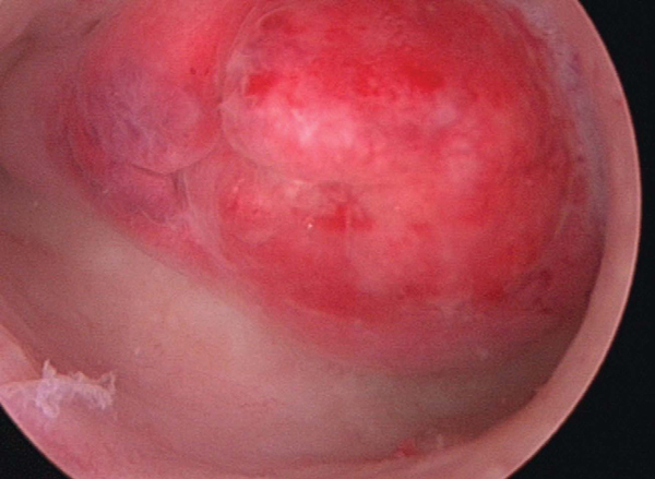
Significant submucous fibroids (see figure 1 above) are also thought to interfere with fertility and implantation. Large fibroids cause pressure symptoms with increasing girth, urinary frequency, constipation and rarely urinary retention. A pedunculated fibroid can occasionally present with torsion.
During pregnancy, fibroids enlarge and may undergo red degeneration causing pain. Large fibroids can cause malpresentation and should be remembered as a rare cause of the unengaged head at term. Because fibroids can interfere with contraction of the uterine muscle, they are associated with postpartum haemorrhage.
Fibroids can occur anywhere in the uterus and are classified according to location:
- Submucous - either partly or wholly within the endometrial cavity
- Intramural - in the muscle within the myometrium
- Subserous - partly within the myometrium and projecting into the pelvis
- Pedunculated - attached to the uterus by a narrow stalk
The exact cause of fibroids is not known. They enlarge when oestrogen levels are high, for example during pregnancy, and shrink when oestrogen levels are low, for example after the menopause. Hormone replacement therapy may cause fibroids to persist after the menopause.
Diagnosis
Fibroids may be found during a routine examination or by chance during a scan for some other reason. On bimanual examination the uterus appears irregular in outline. An ultrasound scan can confirm the diagnosis. If the uterus is >12 weeks in size and/or palpable abdominally, an abdominal scan should be requested. Uterine length in centimetres is equivalent to weeks in size i.e. a 12 cm uterus is equivalent to a 12-week uterus. Women with menorrhagia and fibroids >12 cm and/or a palpable uterus should be referred to a specialist for further opinion.
Symptomatic treatment
If periods are heavy, it may be possible just to manage symptoms until the menopause when fibroids shrink and symptoms improve. There is currently no proven medical treatment to cure fibroids. Medication can only be used to improve symptoms and/or shrink the fibroids prior to surgery.2
The symptomatic treatments listed in table 2 are used to treat heavy periods whatever the cause.
Ulipristal acetate has shown promise in primary research trials.4 Currently used as an emergency contraceptive, it is a progesterone receptor modulator. A randomised double blind placebo controlled trial has concluded that it is effective in shrinking fibroids with few side effects. Further larger clinical trials are needed to establish whether this is a good treatment option.
Surgery
Myomectomy
Women with fibroids >3 cm in diameter causing significant symptoms, pain or pressure and wishing to retain their uterus may consider myomectomy. Abdominal myomectomy is a longer operation than abdominal hysterectomy, but associated with less blood loss and slightly shorter hospital stay (3.96 ± 2.1 versus 4.42 ± 2.4 days).4
Myomectomy leaves a scar on the uterus similar to a midline caesarean section scar which can weaken and/or rupture during pregnancy or childbirth. If the fibroid traverses the hemi-myometrium, and the uterine cavity is entered, caesarean section may be advised as a safer option.
Myomectomy can result in adhesions which may lead to pain and/or impaired fertility. Around one in four women need further surgery, either because more fibroids grow, or because their symptoms have not significantly improved.
Hysterectomy
Hysterectomy is the standard treatment for women with symptomatic fibroids who have not improved with medical treatment. If the woman's family is complete and the fibroids are multiple, hysterectomy provides a permanent cure. Compared with myomectomy, hysterectomy has more blood loss, but the number of women requiring a transfusion is similar. It is normal to conserve healthy ovaries and women also have the option to keep their cervix.
Hysterectomy route is dictated by:
- uterine size
- presence and size of uterine fibroids
- mobility and descent of the uterus
- size and shape of the vagina
- history of previous surgery
Hysteroscopic myomectomy
Fertility is potentially retained with this procedure. If the fibroid is very large or only partially within the endometrial cavity, then it may require more than one operation to resect it. Uncommon side effects are adhesions, which may lead to pain and/or impaired fertility, the need for further surgery, perforation, recurrence or infection. Rarely haemorrhage may occur during surgery and can usually be controlled by insertion of a balloon catheter which is inflated inside the uterine cavity. The catheter can generally be removed a few hours later.
Uterine artery embolisation
This offers a less invasive alternative to hysterectomy or myomectomy and was first introduced in 1991.6 Since then 100,000 procedures have been performed although there are no long-term data available.
The treatment is only recommended if surgery was planned for symptomatic fibroids and if the fibroids are7Uterine artery embolisation is unsuitable for women with pelvic infection, or if the patient would not consent to a hysterectomy under any circumstances.
Treatment is successful in up to nine out of ten women and results in lighter periods, less pain and fewer pressure symptoms. Up to one in ten women undergo a hysterectomy, either because treatment fails or as a result of infection. Death as a result of infection has been reported, particularly in those women with a uterus >20 weeks in size.
The total procedure takes 30 to 90 minutes and is carried out under local anaesthetic given into the skin of the groin. A needle is inserted into the femoral artery and a plastic catheter threaded through the artery to reach the uterine arteries. Small embolisation particles are injected which cause thrombosis and consequent fibroid infarction.
Admission for 24 hours after the procedure is normal because pain can be significant. Other common post-treatment effects are nausea, fever and flu-like symptoms with raised inflammatory markers and white cell count. These effects are usually
self-limiting and are managed by analgesics and anti-inflammatory medications. Readmission may be required in 3-5% of cases for parenteral analgesics and fluid balance control.
MRI guided percutaneous laser ablation
This procedure is carried out under sedation. A local anaesthetic is given into the skin and, under MR image guidance, needles are inserted into the centre of the targeted fibroid.8 Laser fibres are inserted down the centre of each of the needles and laser energy is then used to destroy the fibroid. The area of heating is monitored with the MR scanner. Following treatment, the damaged area forms scar tissue, which contracts causing the fibroid to shrink. Over a year, the average fibroid shrinks by up to 40% with relief of symptoms, especially heavy periods and pressure on the bladder and bowel. This is a very specialised procedure and there is currently only one centre in the UK that undertakes it. As such, the evidence is limited.
Referral
Referral is recommended in the following cases:
- submucous fibroid and abnormal bleeding
- fibroids >3 cm in diameter
- uterus palpable abdominally or >12 cm in size on scan
- persistent intermenstrual bleeding
- age >45 where treatment has failed or been ineffective
- rapidly enlarging fibroids warrant urgent referral
Follow-up
Clinical follow-up is sufficient for most women with fibroids.
Patients who have satisfactory symptom control and are happy with their current treatment can be followed up in primary care. However, patients should be advised to report any new symptoms that develop, particularly enlargement of the fibroids, abnormal bleeding and/or discharge, in which case, urgent referral is required. Sarcomatous change within fibroids is rare, but is normally associated with rapid growth and these women should be referred urgently.
In those patients with large fibroids palpable abdominally, a follow-up in three to six months and then annually to check on the size of the fibroids would be advisable.
Women should be referred where periods are becoming more of a problem and/or are associated with iron deficiency anaemia or they are having to take time off work because of the heaviness of the flow.
REFERENCES
1 Marshall LM, Spiegelman D, Barbieri RL et al. Variation in the incidence of uterine leiomyoma among premenopausal women by age and race. Obstet Gynecol 1997; 90(6):967-73
2 National Institute for Health and Clinical Excellence. CG44. Heavy menstrual bleeding. NICE. London. 2007
3 Department of Reproductive Health, World Health Organization. Medical eligibility criteria for contraceptive use (4th edition). WHO. Geneva. 2010
4 Nieman LK, Blocker W, Nansel T et al. Efficacy and tolerability of CDB-2914 treatment for symptomatic uterine fibroids: a randomized, double-blind, placebo-controlled, phase IIb study. Fertil Steril 2011;95(2):767-72.e1-2. Epub 2010 Nov 5
5 Sawin WS, Pilevsky ND, Berlin JA et al. Comparability of peri-operative morbidity between abdominal myomectomy and hysterectomy for women with uterine leiomyomas. Am J Obst Gynecol 2000;183 (6):1448 -1454
6 Royal College of Obstetricians and Gynaecologists and Royal College of Radiologists. Clinical recommendations on the use of uterine artery embolisation in the management of fibroids (2nd edition). Report of a working party. RCOG Press. London. 2009
7 National Institute for Health and Clinical Excellence. IPG 367. Uterine artery embolisation for fibroids. NICE. London. 2010
8 National Institute for Health and Clinical Excellence. IPG 30. Magnetic Resonance (MR) image-guided percutaneous laser ablation of uterine fibroids. NICE. London. 2003
ARTICLE IN PDF
 Download this article in PDF format
EXTERNAL WEBLINKS
Patient information for women with fibroids has been developed by the Royal Free Hospital, London and can be found at
PubMed
Premature menopause hormone replacement
NICE Evidence
British National Formulary
Treatment of vaginal and vulval conditions
Current evidence - Ovarian cancer
US NCI
PDQ® (Physician Data Query) is NCI's cancer database of peer-reviewed, regularly updated evidence-based, referenced summaries
Ovarian
PubMed
UK NICE
Ovarian
UK Risk Assessment Tools
QCancer
Qcancer women
UK Early diagnosis programmes
UK NCAT
Macmillan - early diagnosis
US NCI
PDQ® (Physician Data Query) is NCI's cancer database of peer-reviewed, regularly updated evidence-based, referenced summaries
Women
Breast
Ovarian
Endometrial screening
Endometrial
Cervical
Preset searches from key journals
British Journal of General Practice
Contraception
HRT
LARC
IUD
British Medical Journal
Contraception
LARC
IUD
HRT
Journal of family planning and reproductive health
LARC
IUD
HRT
New England Journal of Medicine
Contraception
HRT