SEARCH

Ralston S H. Diagnosis and management of osteogenesis imperfecta. Practitioner February 2022;266(1855):11-15
Diagnosis and management of osteogenesis imperfecta
24 Feb 2022
AUTHORS
Professor Stuart H Ralston MD FRCP, Professor of Rheumatology, Rheumatology and Bone Research Group, Centre for Genomic and Experimental Medicine, MRC Institute of Genetics and Cancer, University of Edinburgh, Western General Hospital, Edinburgh, UK
Competing interests: Professor Stuart Ralston is chief investigator on the TOPAZ trial which has received non-financial support from Eli Lilly in the form of a donation of teriparatide as one of the investigational medicinal products. He has received research grants to his institution from Kyowa Kirin, and financial support to his institution for educational events from Pfizer, AbbVie, Kyowa Kirin, Alexion, Amgen, Celgene, Bristol Myers Squibb, Janssen-Cilag, Novartis, Eli Lilly, Thornton & Ross, Astra Zeneca, Sanofi Genzyme, Sandoz and Roche, all outside the submitted work.
Article
Osteogenesis imperfecta is a group of rare inherited skeletal disorders characterised by a greatly increased risk of fragility fractures. The reduction in bone strength is due to defects in its material properties and mineralisation. More than 90% of patients have mutations in either the COL1A1 or COL1A2 genes which are responsible for producing type I collagen, the most abundant protein in bone. Low trauma fractures typically start in infancy e.g. as a consequence of falling when learning to walk. Rarely mild osteogenesis imperfecta can present for the first time during adulthood. Deformity of the rib cage may occur in severe disease, leading to respiratory problems. Patients with severe disease have a marked reduction in mobility and often require a wheelchair. Type I is the most common form and is characterised by the presence of blue sclerae. Other associated features which affect some, but not all, patients with osteogenesis imperfecta are: deafness; dentinogenesis imperfecta (weak, yellow or grey teeth which may chip and break more easily); joint hypermobility; and cardiac disease e.g. mitral or aortic regurgitation. Diagnosis is primarily clinical, based on a history of multiple low trauma fractures during infancy and childhood, and other clinical features.
Osteogenesis imperfecta is a group of rare inherited skeletal disorders characterised by a greatly increased risk of fragility fractures. More than 90% of patients have mutations in either the COL1A1 or COL1A2 genes which are responsible for producing type I collagen, the most abundant protein in bone.
The clinical presentation is with low trauma fractures particularly during infancy and childhood. The increased risk of fracture persists into adulthood and affected individuals can suffer tens or hundreds of fractures during their lifetime.
While bone mineral density (BMD) is low in about one-third of people with osteogenesis imperfecta, the increased risk of fracture is far greater than can be accounted for by reduced BMD.
This indicates that the key mechanism of fracture is reduced bone strength due to defects of bone matrix and mineralisation.
Patients who present to their GP with early onset fractures should be referred to specialist services so that the diagnosis can be confirmed.
Pathophysiology
Osteogenesis imperfecta most commonly results from mutations in either the COL1A1 or COL1A2 genes which encode the type I alpha 1 and type I alpha 2 collagen protein chains. These chains assemble to form type I collagen, a trimer in which the individual protein chains are wound together in a triple helix.
The disease-causing mutations can either alter the amino acid sequence, which interferes with assembly of the individual protein chains into the triple helix (qualitative mutations) or can reduce the absolute amount of collagen being produced (quantitative mutations).
At a molecular level, quantitative mutations introduce premature stop codons into the RNA molecule resulting in nonsense mediated RNA degradation, which eliminates the abnormal RNA but results in a 50% reduction in the amount of protein produced. In general, qualitative mutations cause a more severe phenotype than quantitative mutations.
Less commonly, mutations in other genes can be responsible (see table 1). Many of these are involved in modifying the collagen proteins after they are produced whereas others affect the function of bone forming cells.
It is beyond the scope of this article to go into detail on disease mechanisms for all causal genes but readers who may wish to learn more are directed to recent review articles on this topic.1,2
The result of these abnormalities of bone matrix is to reduce bone strength, which accounts for multiple fractures that are the hallmark of the disease.
Although fractures occur in osteoporosis where bone fragility is also increased, the two conditions are fundamentally different. The main problem in osteogenesis imperfecta is a reduction in the strength of bone due to defects in its material properties and mineralisation. In osteoporosis, bone matrix and mineralisation are normal but are reduced in amount.
Mode of inheritance
Osteogenesis imperfecta can be inherited in an autosomal dominant or autosomal recessive pattern. Those with mild disease have an autosomal dominant transmission such that one in two individuals in a family are affected in successive generations.
Severe disease is often inherited recessively, and in this case there may be consanguinity and a previously affected family member.
A family history is not always present either because of a new mutation or parental mosaicism where the mutation is present in a proportion of the parents’ germ cells.
It has been estimated that around 25% of individuals with osteogenesis imperfecta are born into a family where there is no previous history of the disorder.
An X-linked dominant condition has been described with similarities to osteogenesis imperfecta caused by mutations in the PLS3 gene.3 This is considered to be a form of early onset osteoporosis rather than osteogenesis imperfecta but is mentioned here as it also presents with severe bone fragility and fractures in childhood.
Epidemiology
The prevalence of osteogenesis imperfecta in the UK has not been studied but population studies in Denmark have given an estimated prevalence of around 1 in 10,000 individuals.4 The Brittle Bone Society, a patient support group, has estimated that there are about 5,000 individuals living with osteogenesis imperfecta in the UK. (https://brittlebone.org)
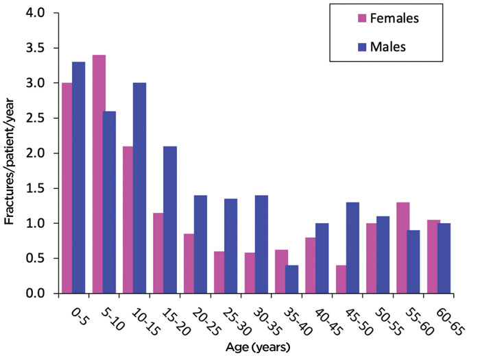
Osteogenesis imperfecta occurs in both sexes and affects all ethnic groups. Fractures occur most commonly in childhood and fall in incidence thereafter with a higher prevalence in males during adolescence and adulthood.5 (see figure 1).
The reduction in fractures during late adolescence and adulthood has been attributed to the increase in bone mass as the skeleton matures and behaviour changes with avoidance of activities that place the individual at risk of fractures.
Even though the rates decrease during adulthood, fractures occur between 10 and 30 times more frequently in adults with osteogenesis imperfecta than the general population.5
Life expectancy is reduced in osteogenesis imperfecta as compared with the general population, with an increased risk of death as a result of respiratory and gastrointestinal diseases and trauma.6
Clinical features
Osteogenesis imperfecta presents with low trauma fractures as the result of what would normally be trivial injuries typically starting in infancy, e.g. falling when learning to walk. Rarely mild osteogenesis imperfecta can present for the first time during adulthood.
The Sillence classification, which was devised in 1979,7 divided patients with osteogenesis imperfecta into four subtypes (I-IV) based on clinical severity, ranging from mild to lethal.
As new genes for osteogenesis imperfecta have been discovered an extended classification system has been suggested1 which introduces new subtypes related to the underlying genetic abnormality while retaining the Sillence classification for defects associated with mutations in the type I collagen genes (see table 1).
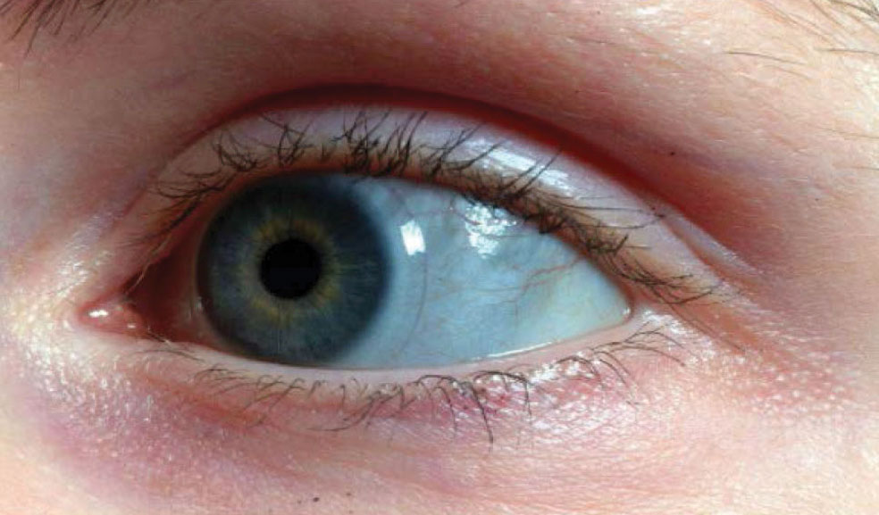
Type I osteogenesis imperfecta is the most common form and is characterised by the presence of blue sclerae (see figure 2) which is caused by a reduced amount of type I collagen in the sclera.8 Some patients with osteogenesis imperfecta also develop deafness due to involvement of bones in the middle ear.
Patients may exhibit grey or yellow discoloration of their teeth; so called dentinogenesis imperfecta, where the teeth may chip and break more easily.8,9
Extraskeletal problems also occur since type I collagen is not only present in bone, but in other tissues as well. Mitral regurgitation, aortic regurgitation, atrial fibrillation and heart failure are more common in osteogenesis imperfecta than the general population.10
Ligament laxity, tendon rupture and joint hypermobility are recognised features of the disease.11 Osteoarthritis can also occur but it is not clear if this is more frequent in osteogenesis imperfecta than in the general population.
Patients with osteogenesis imperfecta commonly exhibit bone deformity, scoliosis and kyphosis due to vertebral fractures. Such deformities are invariably present in severe forms of osteogenesis imperfecta (Sillence type III and IV) but can also occur in patients with milder disease (type I).
Deformity of the rib cage may occur in severe disease leading to respiratory problems. Patients with severe disease have a marked reduction in mobility and often require a wheelchair to gain mobility and independence during childhood, adolescence and beyond.
Diagnosis
The diagnosis of osteogenesis imperfecta is primarily clinical based on a history of multiple low trauma fractures during infancy and childhood, along with other clinical features discussed above.
Genetic testing is being increasingly used for diagnosis; to clarify the molecular basis of the disease and for genetic counselling. Genetic testing for osteogenesis imperfecta can be done by referral to clinical genetics services either directly or through specialised clinics for patients with metabolic bone disease.
Non-accidental injury may enter into the differential diagnosis. Features that would lead one to suspect non-accidental injury are the absence of other clinical features of osteogenesis imperfecta, bilateral rib fractures, skull fractures, metaphyseal fractures and bruising at sites distant from fractures.12 Any of these features should prompt specialist referral.
Management
The management of osteogenesis imperfecta is multidisciplinary in nature and the multidisciplinary team (MDT) is usually coordinated by a physician with expertise in metabolic bone disease.13 This individual will make the initial diagnosis, decide upon the need for medical therapy and refer onwards to other members of the MDT when required. Individual management components are discussed below.
Medical management
There is no licensed medical treatment for the prevention of fractures in osteogenesis imperfecta or for the treatment of other features of the disease. Intravenous bisphosphonates including pamidronate and zoledronic acid are widely used in children with osteogenesis imperfecta. However, the evidence that these treatments reduce the risk of fracture is weak, largely based on observational studies.14
Randomised placebo controlled trials have yielded conflicting results and a meta-analysis of these trials has shown limited evidence of benefit.15 Despite the weak evidence base, these medicines are used widely in paediatric specialist units and are considered to be standard of care in children with osteogenesis imperfecta. Observational studies have suggested that intravenous bisphosphonate therapy might help to modify progression of scoliosis in type lll but did not seem to offer benefit in other forms of osteogenesis imperfecta.16
Concomitant treatment with calcium and vitamin D supplements may be required to mitigate against hypocalcaemia in those treated with parenteral bisphosphonates.
Bisphosphonates are used much less frequently in adults with osteogenesis imperfecta since there is a concern that long-term treatment may increase the risk of fractures by oversuppressing bone remodelling.
Various other treatments used in osteoporosis, including denosumab and teriparatide, have been explored but have not gained widespread acceptance in routine clinical practice. Supportive therapies may be required to control pain associated with bone deformities and vertebral fractures.
Occupational therapy
Occupational therapy plays an important role, particularly in individuals with more severe forms of osteogenesis imperfecta. The occupational therapist will assess the patient and advise on devices and mobility aids to allow affected individuals to optimise function.
Physiotherapy
The physiotherapist can advise the parents of infants and children on safe handling as well as helping to set goals for reaching developmental milestones. One of the most important goals is to help the child gain independence in terms of mobility either using walking aids or a wheelchair.
Physiotherapy supervised exercise programmes can positively influence muscle strength and physical ability.17,18
Anecdotal evidence suggests that improving muscle strength can also have beneficial effects in stabilising joints in those with hypermobility.
In previous years, people with osteogenesis imperfecta were discouraged from participating in sports but there are now many examples of individuals with osteogenesis imperfecta reaching a high level in sporting activities. This in turn can have beneficial effects on wellbeing and quality of life.
Orthotics
Ankle foot orthoses can control the position of the distal lower extremity and may have a role in preventing the worsening of foot deformity. Removable splints for the upper limb can be used to help ambulation in supporting patients who use a self-propelled wheelchair.
An individualised approach to the use of splints is required and an experienced orthotist can be invaluable for these patients.
Orthopaedic surgery
Orthopaedic surgery plays a central role not only in the management of fractures but also as prophylactic surgery to prevent and correct deformities. The aims of orthopaedic management are to maximise function and minimise pain. Children with osteogenesis imperfecta tend to become very well known to their local A&E and orthopaedic services because of their recurrent fractures.
A conservative approach is usually taken and fixation rarely performed in the acute setting. Splinting and casting is normally used and after a number of fractures, the parents and patients themselves become very adept at applying casts and may do this at home reducing the need for hospital visits.
Early mobilisation is encouraged since repeated spells of non-weightbearing lead to muscle wasting and loss of the ability to ambulate independently.
Management of fractures in adults can be challenging due to the presence of deformity, previous surgery and/or metalwork.19
Surgery to correct limb deformity may be required for a patient to remain ambulant. Technical details of the various approaches that may be used to correct limb deformities is beyond the scope of this article, but this topic has been the subject of a recent review.20
Spinal surgery may also be indicated to correct scoliosis but this would generally require referral to a tertiary centre with experience in this area.21
Other MDT input
The input of other healthcare professionals including clinical psychologists, speech and language therapists, dietitians, and social workers also has an important role.
Future prospects
There have been major strides in understanding the genetic basis of osteogenesis imperfecta over recent decades, but this has not been matched by improvements in management.
This is due in part to the lack of appropriately powered clinical trials to examine the effects of therapeutic interventions on fractures which are the main cause of morbidity. Such trials are now in progress and it is hoped that they will yield some useful information over the next five years.
Exploratory studies are also being performed to look at the feasibility of stem cell therapy, and genetic approaches such as RNA silencing and gene editing with CRISPR/Cas to improve outcomes in this disabling disease.
Competing interests: Professor Stuart Ralston is chief investigator on the TOPAZ trial which has received non-financial support from Eli Lilly in the form of a donation of teriparatide as one of the investigational medicinal products. He has received research grants to his institution from Kyowa Kirin, and financial support to his institution for educational events from Pfizer, AbbVie, Kyowa Kirin, Alexion, Amgen, Celgene, Bristol Myers Squibb, Janssen-Cilag, Novartis, Eli Lilly, Thornton & Ross, Astra Zeneca, Sanofi Genzyme, Sandoz and Roche, all outside the submitted work.
REFERENCES
1 Forlino A, Marini JC. Osteogenesis imperfecta. Lancet 2016;387:1657-71
2 Marom R, Lee YC, Grafe I, Lee B. Pharmacological and biological therapeutic strategies for osteogenesis imperfecta. Am J Med Genet C Semin Med Genet 2016;172:367-83
3 van Dijk FS, Zillikens MC, Micha D et al. PLS3 mutations in X-linked osteoporosis with fractures. N Engl J Med 2013;369:1529-36
4 Andersen PE Jr, Hauge M. Osteogenesis imperfecta: a genetic, radiological, and epidemiological study. Clin Genet 1989;36:250-55
5 Folkestad L, Hald JD, Ersboll AK et al. Fracture rates and fracture sites in patients with osteogenesis imperfecta: a nationwide register-based cohort study. J Bone Miner Res 2017;32:125-34
6 Folkestad L. Mortality and morbidity in patients with osteogenesis imperfecta in Denmark. Dan Med J 2018;65(4):B5454
7 Sillence DO, Senn A, Danks DM. Genetic heterogeneity in osteogenesis imperfecta. J Med Genet 1979;16:101-16
8 Hald JD, Folkestad L, Swan CZ et al. Osteogenesis imperfecta and the teeth, eyes, and ears-a study of non-skeletal phenotypes in adults. Osteoporos Int 2018;29:2781-89
9 Retrouvey JM, Schwartz S, Hartsfield JK et al. Oral-facial aspects of osteogenesis imperfecta. In: Osteogenesis imperfecta - a translational approach to brittle bone disease. Elsevier. New York. pp 313-27
10 Folkestad L, Hald JD, Gram J et al. Cardiovascular disease in patients with osteogenesis imperfecta - a nationwide, register-based cohort study. Int J Cardiol 2016;225:250-57
11 McKiernan FE.Musculoskeletal manifestations of mild osteogenesis imperfecta in the adult. Osteoporos Int 2005;16:1698-1702
12 Pereira EM. Clinical perspectives on osteogenesis imperfecta versus non-accidental injury. Am J Med Genet C Semin Med Genet 2015;169(4):302-06
13 Marr C, Seasman A, Bishop N. Managing the patient with osteogenesis imperfecta: a multidisciplinary approach. J Multidiscip Healthc 2017;10:145-55
14 Glorieux FH, Bishop NJ, Plotkin H et al. Cyclic administration of pamidronate in children with severe osteogenesis imperfecta. N Engl J Med 1998;339:947-52
15 Hald JD, Evangelou E, Langdahl BL, Ralston SH. Bisphosphonates for the prevention of fractures in osteogenesis imperfecta: meta-analysis of placebo-controlled trials. J Bone Miner Res 2015;30:929-33
16 Sato A, Ouellet J, Muneta T et al. Scoliosis in osteogenesis imperfecta caused by COL1A1/COL1A2 mutations - genotype-phenotype correlations and effect of bisphosphonate treatment. Bone 2016;86:53-57
17 Binder H, Conway A, Gerber LH. Rehabilitation approaches to children with osteogenesis imperfecta: a ten-year experience. Arch Phys Med Rehabil 1993;74:386-90
18 Binder H, Conway A, Hason S et al. Comprehensive rehabilitation of the child with osteogenesis imperfecta. Am J Med Genet 1993;45:265-69
19 Gil JA, DeFroda SF, Sindhu K et al. Challenges of fracture management for adults with osteogenesis imperfecta. Orthopedics 2017;40:e17-e22
20 Ralston SH, Gaston MS. Management of osteogenesis imperfecta. Front Endocrinol (Lausanne) 2019;10:924
21 Puvanesarajah V, Sonseller PD. Scoliosis and kyphosis in osteogenesis imperfecta. In: Osteogenesis imperfecta - a translational approach to brittle bone disease. Shapiro JR, Byers PH, Glorieux FH, Sponseller PD (eds). Academic Press. New York. 2014. Chapter 43, pp 401-06
 Download this article in PDF format