SEARCH

Sinnappurajar P, Ramanan AV. Juvenile spondyloarthropathies often go unrecognised. Practitioner June 2021;265(1849):11-15
Juvenile spondyloarthropathies often go unrecognised
25 Jun 2021
AUTHORS
Dr Praisoody Sinnappurajar MBBS DCH, Clinical Fellow in Paediatric Rheumatology
Professor Athimalaipet V Ramanan FRCPCH FRCP, Consultant Paediatric Rheumatologist
Bristol Royal Hospital for Children, University Hospitals Bristol NHS Foundation Trust, Bristol, UK
Competing interests
Professor Athimalaipet Ramanan has received speaker and consulting fees from AbbVie, Eli Lilly, Roche, Novartis, UCB and Sobi.
Dr Praisoody Sinnappurajar has no competing interests.
Article
Juvenile spondyloarthropathies (JSpA) are a heterogeneous group of inflammatory disorders that present in children under the age of 16 years. Unlike other arthritides, they are not defined by the presence of rheumatoid factor (RF) or antinuclear antibody (ANA), but are associated with the presence of HLA-B27 in up to 50% of JSpA patients, signalling a role in the pathogenesis of JSpA. As the symptoms of JSpA differ from those of adult spondyloarthropathy, diagnosis can be challenging. JSpA is an umbrella term encompassing enthesitis-related arthritis (ERA), juvenile onset ankylosing spondylitis (JoAS), juvenile psoriatic arthritis (JPsA), IBD-related arthritis and reactive arthritis. Initial assessment of any childhood arthropathy should include a full blood count with differentials and inflammatory markers (CRP and ESR), which will be normal in most cases of JSpA, or may reflect a mild inflammatory response and anaemia relating to chronic disease. RF and ANA are characteristically negative. Children with JSpA should be tested for HLA-B27. A faecal calprotectin test may be considered if there are symptoms of gut inflammation. Children with suspected JSpA should be referred via their GP to a tertiary paediatric rheumatology centre for specialist multidisciplinary input from a team including paediatric rheumatologists, paediatric ophthalmologists, specialist nurses, physiotherapists, occupational therapists and psychologists. The goals of therapy include preventing joint damage and achieving remission. Uveitis should be urgently referred for specialist ophthalmology review.
Juvenile spondylo-arthropathies (JSpA) are a heterogeneous group of inflammatory disorders that present in children under the age of 16 years.
JSpA is an umbrella term encompassing enthesitis-related arthritis (ERA), juvenile onset ankylosing spondylitis (JoAS), juvenile psoriatic arthritis (JPsA), inflammatory bowel disease (IBD)-related arthritis and reactive arthritis.1 The prevalence of ERA and JPsA in Europe is estimated to be up to 6.5 per 100,000 population under the age of 16.2
Unlike other arthritides, they are not defined by the presence of rheumatoid factor (RF) or antinuclear antibody (ANA), but are associated with the presence of HLA-B27. This molecule is found in up to 50% of JSpA patients, signalling a role in the pathogenesis of JSpA.3
As the symptoms of JSpA differ from those of adult spondyloarthropathy, diagnosis can be challenging. Up to 18% of cases of ankylosing spondylitis can begin in adolescence, but there is a mean interval from symptom onset to formal diagnosis of eight years. These symptoms may be indolent or can be mistaken for ‘growing pains’, which might lead families to delay in seeking a medical opinion.4 GPs play an important role in the detection, diagnosis and management of children in this under recognised group.
Classification
There are a number of proposed classification criteria for juvenile arthritis, none of which specifically address JSpA.
The 2001 revision of the International League of Associations for Rheumatology (ILAR) classification, which proposes seven different categories of juvenile idiopathic arthritis (JIA), is the most commonly used.5 Using this system, most cases of JSpA will be classified as ERA or JPsA, or undifferentiated arthritis (see table 1).
There are several limitations to applying the ILAR criteria to JSpA including the lack of specific categories for IBD-related arthritis or a reactive arthritis, which is generally a peripheral arthritis with onset within six weeks of an infection. Similarly, JoAS is not specifically referenced in the ILAR categories.
Some researchers argue that ERA and JoAS exist on the same spectrum, with the former progressing to the latter.6,7,8 The same criteria used for diagnosing adult ankylosing spondylitis (AS), such as the New York modified criteria, can be applied to children to diagnose JoAS.7 For the purpose of this article, JSpA are considered as an entity.
Clinical features
JSpA disorders occur more commonly in adolescent males, with a large cohort study of ERA demonstrating a 9:1 male predominance and a mean age at diagnosis of 11.3 years.9
The symptoms differ from adult onset spondyloarthropathies. Children tend to present with a peripheral arthritis or enthesitis, although some may present with inflammatory back pain. Clinical features of JSpA are listed in table 2.
Previous studies have reported the mean interval between onset of peripheral arthritis and inflammatory back pain in JoAS as 5.5 years, which demonstrates the continuum that exists in JSpA.10 The challenge is to identify these patients early and refer them to a paediatric rheumatologist for assessment.
Systemic symptoms such as fever or fatigue are typically absent in JSpA and should prompt investigation for another diagnosis.11 Other features in the history to note include any recent symptoms of infection (e.g. rhinorrhoea, cough, diarrhoea), which might suggest a reactive arthritis, and whether there is a relevant family history.
Enthesitis
Enthesitis, inflammation at the attachment point of tendons or ligaments to bone, is a distinct feature of JSpA, more frequently seen in children than in adults. Typically, the enthesitis will be multifocal, symmetrical and persistent. It most commonly affects the plantar fascia and Achilles tendon attachments on the heel, and the tendon attachments on the patella (at 2, 6 and 10 o’clock positions).3
Careful examination of all frequently affected entheseal insertion sites, by applying pressure with the thumb until the nailbed blanches, should elicit localised tenderness. Gait examination might reveal an avoidance to weight bear on certain entheses.
The history should correlate with clinical findings, as entheseal pain can also be seen in otherwise healthy children, as well as in overuse injuries and Osgood-Schlatter’s disease.12
Peripheral arthritis
The presenting arthritis of JSpA is typically peripheral, oligoarticular (< 5 joints), asymmetric, and involves the large joints of the lower extremities (hips, knees, ankles). The patient might report morning stiffness or joint pain that improves with activity.
Examination can reveal joint swelling, pain on passive movement and a restricted range of movement in the affected joint. The paediatric Gait, Arms, Legs, Spine (pGALS) is a useful framework to guide examination.
Tarsitis, inflammation of the midfoot, is most commonly seen in the JSpA disorders, while dactylitis, sausage-like inflammation of the digits, is typical of psoriatic arthritis.1,13
Axial arthritis
Unlike patients with adult spondyloarthropathies, symptomatic axial involvement (spine, sacroiliac joints) is less common at disease onset, but should be monitored for, as it can evolve later. A retrospective study of 114 patients with JSpA demonstrated that axial involvement increased from 35% of patients at baseline to 63% by five years’ follow-up.14 The diagnosis of sacroiliitis in children remains controversial, as it can be clinically silent, but radiologically evident, in up to 20% of JSpA patients.13
Typical clinical features that suggest sacroiliitis are outlined in table 3, above, and can include alternating buttock pain, or chronic back pain and stiffness. Left untreated, sacroiliitis may progress to spondylitis, inflammation of the vertebral bones, or ankylosis (fusion).15
Early aggressive intervention in children with active sacroiliitis is recommended as per the American College of Rheumatology guidelines.16
Extra-articular manifestations
GPs should have a high index of suspicion in JSpA patients with a painful red eye, as acute anterior uveitis can occur in up to 25% of children with JSpA. The uveitis of JSpA is typically acute in onset, unilateral, with associated pain, conjunctival injection and photophobia.3 This is in contrast to other forms of JIA, which are associated with chronic uveitis that may be asymptomatic until loss of vision occurs.
Large cohort studies in adult populations have found that 40-50% of patients presenting with idiopathic acute anterior uveitis have an underlying spondyloarthritis.17,18
Patients presenting with uveitis should be urgently referred for specialist ophthalmology review.19
JSpA is found in approximately 10% of children with IBD, more often in Crohn's disease than ulcerative colitis.
Arthritis most commonly follows the onset of enteropathy. Features of gut inflammation, such as abdominal pain, diarrhoea, bloody stools and poor growth, are therefore pertinent.20
Skin manifestations are most commonly seen with psoriatic arthritis, including the typical scaling plaques on the hairline, behind the ears, on the elbows and knees. Other changes of psoriasis include nail pitting and onycholysis.
Uncommonly, aortic insufficiency, myocarditis, endocarditis and pericarditis can occur in JSpA patients, often with spontaneous resolution.20
Investigations
Although no single test can diagnose JSpA, certain blood tests and imaging may provide important clues. Initial assessment of any childhood arthropathy should include a full blood count with differentials and inflammatory markers (CRP and ESR), which will be normal in most cases of JSpA or may reflect a mild inflammatory response and anaemia relating to chronic disease.
RF and ANA are characteristically negative. Children with JSpA should be tested for HLA-B27. A faecal calprotectin test may be considered if there are symptoms of gut inflammation, and levels are typically higher in JSpA (ERA) than other forms of JIA.8
Various imaging modalities can be considered. Plain radiographs of the joint in question may assist in excluding malignancy or trauma. Radiographs may also be utilised in the assessment of sacroiliitis, although their interpretation in children is challenging and will only reveal changes after some degree of joint destruction has occurred.
Ultrasound may confirm synovitis or subtle effusions in peripheral joints, and can also be useful in the assessment of clinical or subclinical enthesitis.13
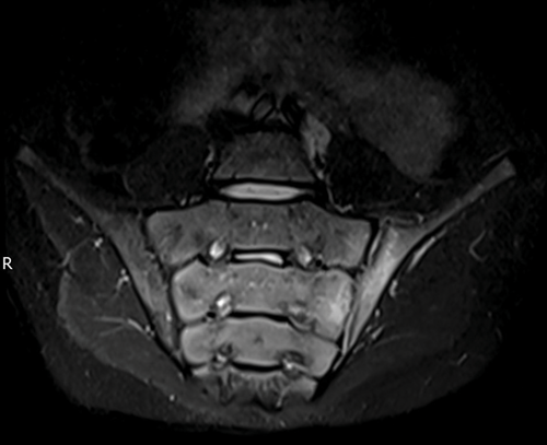
MRI is more sensitive than other modalities in detecting sacroiliitis (see figure 1) revealing active inflammation in 42% of children with JSpA with normal radiographs. However, in the absence of axial signs or symptoms, it can be difficult to know when to proceed to such imaging in children with JSpA.22
The presence of sacroiliitis is an important differentiator for prognostication and treatment intensity, and further guidelines are required to understand when imaging should be pursued.
Management
Children with suspected JSpA should be referred via their GP to a tertiary paediatric rheumatology centre for specialist multidisciplinary team input from paediatric rheumatologists, paediatric ophthalmologists, specialist nurses, physiotherapists, occupational therapists and psychologists.
The goals of therapy include preventing joint damage and achieving remission while balancing pharmacological burden and side effects.8
The absence of a centralised classification system has inhibited a unified treatment approach. The 2019 American College of Rheumatology guidelines for the treatment of JIA approach their recommendations from a practice perspective, suggesting treatment based on the number of active joints and the presence of sacroiliitis, rather than on the specific subtype of JIA.16
Ideally, only NSAIDs should be instituted in a primary care setting. Corticosteroids, disease modifying anti-rheumatic drugs (DMARDs) and biologics should be initiated by a specialist paediatric rheumatologist.
NSAIDs
NSAIDs are a first-line therapy that can provide both anti-inflammatory and analgesic effects and may be initiated by GPs in children with clinical arthritis or enthesitis, while awaiting formal specialist review. NSAID monotherapy should be ceased after two months if it does not induce disease remission. Naproxen is generally the preferred NSAID, as it has a convenient twice daily regimen. Toxicity monitoring includes full blood count, creatinine, liver function testing and urinalysis.8
Corticosteroids
Oral corticosteroids may be instituted for short-term respite of peripheral arthritis but are not a mainstay of JSpA treatment.
Systemic steroids have a number of adverse effects in the acute setting, including insomnia, mood changes, weight gain, hypertension and hyperglycaemia, which should be monitored.3
Generally, a paediatric rheumatologist might consider a course of steroids as a bridging therapy if there are ongoing symptoms or relapse.8
Intraarticular (IA) administration of steroids may also be used, depending on the age of the child, number of joints involved and the sites. IA steroids can help rapidly relieve symptoms within days and avoid the need for systemic steroids.8
DMARDs
DMARDs, such as methotrexate and sulfasalazine, may be used in a purely peripheral arthritis. However, they have not shown efficacy for axial disease or enthesitis and biologic agents are usually the preferred choice if these features are present. Sulfasalazine and methotrexate have an adverse effect profile including bone marrow suppression, nephrotoxicity and hepatotoxicity.8 The specialist team will communicate the plan for monitoring to both the patient and their GP.
Biologic agents
Tumour necrosis factor (TNF) inhibitors, such as adalimumab, infliximab and etanercept, have demonstrated efficacy in arthritis, enthesitis, and the symptomatic treatment of axial disease in JSpA.
In adults, delay in initiation of TNF inhibitors has been associated with increased risk of progressive structural damage.22 They should therefore be considered early in the course of axial disease, as well as in a peripheral arthritis associated with JSpA, refractory to the above.
The 2019 American College of Rheumatology recommendations suggest adding a TNF inhibitor in children with ongoing active sacroiliitis despite NSAID therapy, bypassing DMARDs, and considering oral steroids only as bridging therapy.16
The potential adverse effects of TNF inhibitors include predisposition to infection, activation of latent tuberculosis, cytopenia, hypersensitivity reactions, as well as psoriasis, demyelination and malignancy.
In addition, biologics may become ineffectual over time due to the development of human antichimeric antibodies.23
Other biologic agents, such as the interleukin (IL)-6 inhibitor, tocilizumab, commonly used in other forms of JIA, have not demonstrated efficacy in JSpA.8
Prospective therapeutic agents
There is increasing evidence for the role of interleukins IL-17 and IL-23 in the pathogenesis of spondyloarthropathies. Biologic inhibitors targeting these cytokines, such as secukinumab (IL-17) and ustekinumab (IL-12, IL-23), have been successfully used in trials of adult patients with spondyloarthritis.24 Both biologics are currently licensed in the UK for psoriatic arthritis in adults.
The results of a two-year multicentre randomised, double-blind, placebo-controlled trial assessing secukinumab in JPsA and ERA which show a significant delay in time to flare vs placebo, and a favourable safety profile, are due to be released in the near future.25
Additionally, the janus kinase (JAK) inhibitor tofacitinib has been approved for use in adults with psoriatic arthritis in the UK, and may hold promise for use in children in the future, with the added benefit of oral administration.24
Monitoring
All children with JSpA should maintain active follow-up with both their GP and a paediatric rheumatology service. The GP plays a key role in facilitating medication toxicity monitoring, as well as communicating with the specialist team if the patient experiences a flare. Monitoring of disease activity in JSpA may be undertaken using the JSpA disease activity score, but this varies between centres.25 Regular blood tests are required to monitor medication toxicity, but generally will not indicate disease flare or quiescence.
Families of children with JSpA should be educated regarding the warning signs for uveitis to ensure they seek prompt review.4
Despite its important treatment implications, there remains no consensus on early screening for sacroiliitis.28
Conclusion
JSpA generally has a poorer prognosis than other types of JIA. In some studies, fewer than 20% of children with JSpA achieve remission within five years of diagnosis, with the presence of HLA-B27 being a particularly poor prognostic factor.1,9
Sacroiliitis, the hallmark red flag of these disorders, remains poorly defined in children, and further studies regarding its natural history in JSpA is required.
A validated international classification system is needed to facilitate understanding and research, as well as guide standardised management.
The future for children with JSpA, however, appears optimistic, with the promise of targeted biologic agents.
REFERENCES
1 Weiss PF, Colbert RA. Juvenile spondyloarthritis. Pediatr Clin North Am 2018;65(4):675-90
2 Schanberg LE, Ramanan AV, De Benedetti F et al. Toward accelerated authorization and access to new medicines for juvenile idiopathic arthritis. Arthritis Rheumatol 2019;71(12):1976-84
3 Gmuca S, Weiss PF. Juvenile spondyloarthritis. Curr Opin Rheumatol 2015;27(4):364-72
4 Jadon DR, Shaddick G, Jobling A et al. Clinical outcomes and progression to orthopedic surgery in juvenile- versus adult-onset ankylosing spondylitis. Arthritis Care Res 2015;67(5):651-57
5 Petty R, Southwood T, Manners P et al. International League of Associations for Rheumatology Classification of Juvenile Idiopathic Arthritis: Second Revision, Edmonton, 2001. J Rheumatol 2004;31:390-92
6 Elyan M, Khan MA. Diagnosing ankylosing spondylitis. J Rheumatol Suppl 2006;78:12-23
7 Colbert RA. Classification of juvenile spondyloarthritis: enthesitis-related arthritis and beyond. Nat Rev Rheumatol 2010;6(8):477-85
8 Hahn Y-S. Enthesitis-related arthritis. J Rheum Dis 2018;25(4):221-30
9 Gmuca S, Xiao R, Brandon TG et al. Multicenter inception cohort of enthesitis-related arthritis: variation in disease characteristics and treatment approaches. Arthritis Res Ther 2017;19(1):84
10 Jadon DR, Ramanan AV, Sengupta R. Juvenile versus adult-onset ankylosing spondylitis — clinical, radiographic, and social outcomes. A systematic review. J Rheumatol 2013;40(11):1797-1805
11 Mistry RR, Patro P, Agarwal V et al. Enthesitis-related arthritis: current perspectives. Rheumatol Res Rev 2019;11:19-31
12 Ramanathan A, Srinivasalu H, Colbert RA. Update on juvenile spondyloarthritis. Rheum Dis Clin North Am 2013;39(4):767-88
13 Sudoł-Szopińska I, Gietka P, Znajdek M et al. Imaging of juvenile spondyloarthritis. Part I: classifications and radiographs. J Ultrason 2017;17(70):167-75
14 Goirand M, Breton S, Chevallier F et al. Clinical features of children with enthesitis-related juvenile idiopathic arthritis/juvenile spondyloarthritis followed in a French tertiary care pediatric rheumatology centre. Pediatr Rheumatol 2018;16(1):21
15 Weiss PF. Diagnosis and treatment of enthesitis-related arthritis. Adolesc Health Med Ther 2012;3:67-74
16 Ringold S, Angeles-Han ST, Beukelman T et al. 2019 American College of Rheumatology/Arthritis Foundation guideline for the treatment of juvenile idiopathic arthritis: therapeutic approaches for non-systemic polyarthritis, sacroiliitis, and enthesitis. Arthritis Care Res 2019;71(6):717-34
17 Juanola X, Santamaria EL, Condero-Coma M. Description and prevalence of spondyloarthritis in patients with anterior uveitis. Ophthalmology 2016;123(8):1623-36
18 Haroon M, O’Rourke M, Ramasamy P, Murphy C et al. A novel evidence-based detection of undiagnosed spondyloarthritis in patients presenting with acute anterior uveitis: the DUET (Dublin Uveitis Evaluation Tool). Ann Rheum Dis 2015;74:1990-95
19 Marino A, Weiss PF, Brandon TG et al. Juvenile spondyloarthritis: focus on uveitis. Pediatr Rheumatol 2020;18(1):70
20 Sudoł-Szopińska I, Eshed I, Jans L et al. Classifications and imaging of juvenile spondyloarthritis. J Ultrason 2018;18(74):224-33
21 Pagnini I, Scavone M, Maccora I et al. The development of extra-articular manifestations in children with enthesitis-related arthritis: natural course or different disease entity? Front Med 2021; 8 [Last accessed May 23 2021]
22 Sudoł-Szopińska I, Znajdek M, Gietka P et al. Imaging of juvenile spondyloarthritis. Part II: ultrasonography and magnetic resonance imaging. J Ultrason 2017;17(70):176-81
23 Gmuca S, Weiss PF. Evaluation and treatment of childhood enthesitis-related arthritis. Curr Treat Options Rheumatol 2015;1(4):350-64
24 Marino A, Souza MD, Giani T et al. Pharmacotherapy for juvenile spondyloarthritis: an overview of the available therapies. Expert Opin Pharmacother 2020;21(17):2161-68
25 Ruperto N, Foeldvari I, Alexeeva E et al. LB0004 efficacy and safety of secukinumab in enthesitis-related arthritis and juvenile psoriatic arthritis: primary results from a randomised, double-blind, placebo-controlled, treatment withdrawal, phase 3 study (Junipera). Ann Rheum Dis 2021;80:201-02
26 Zanwar A, Phatak S, Aggarwal A. Prospective validation of the juvenile spondyloarthritis disease activity index in children with enthesitis-related arthritis. Rheumatology 2018;57(12):2167-71
27 Angeles‐Han ST, Ringold S, Beukelman T et al. 2019 American College of Rheumatology/Arthritis Foundation guideline for the screening, monitoring, and treatment of juvenile idiopathic arthritis-associated uveitis. Arthritis Care Res 2019;71(6):703-16
28 Weiss PF, Xiao R, Biko DM et al. Sacroiliitis at diagnosis of juvenile spondyloarthritis assessed by radiography, magnetic resonance imaging, and clinical examination. Arthritis Care Res 2016 ;68(2):187-94
 Download this article in PDF format