SEARCH

The Practitioner 2010; 254 (1732): 19–23
Systematic approach needed to establish cause of vertigo
20 Sep 2010
AUTHORS
Dr Ben Turner MBBS MRCP DM
Consultant neurologist and honorary senior lecturer,
Barts & The London NHS Trust
Mr Nicholas Eynon-Lewis BSc (Hons) FRCS Consultant ENT surgeon,
Barts & The London NHS Trust and The Homerton University Hospital
Article
Vertigo is a common but complex challenge. In one postal survey of patients from general practice, 7% reported at least one episode of vertigo in the past year.1 A full-time GP may expect to encounter 10-20 cases of vertigo each year2 and it is often seen as a potentially difficult symptom to diagnose and manage.
The first problem is that the terms vertigo, dizziness, giddiness and imbalance are used in different ways by different clinicians. Dizziness and vertigo are often used interchangeably by both patients and clinicians (including the authors).
The second problem is that the control of balance is complicated and diagnosing the cause of a balance disorder can take time which is usually at a premium in most clinics. Third, vertigo can be caused by many different pathologies, some of which are potentially life threatening, and it is managed by several specialties.
However, much of this can be overcome by using a logical, systematic approach to the diagnosis and subsequent referral or management of the patient.
Vertigo can be defined as an illusion or hallucination of movement. Although the word is derived from the latin vertere which implies a sense of turning, most clinicians would classify vertigo as being rotatory or non rotatory.
Although rotatory vertigo is usually caused by an abnormality of the peripheral vestibular system it can also be seen in central vestibular and other neurological conditions. Chronic vertigo which is not episodic tends to be associated with a sense of instability or sway and is not rotatory. Ataxia is an abnormality of the co-ordination of muscle movement and should not be confused with vertigo.
Maintaining balance is complicated although a basic knowledge of the anatomy and physiology can be extremely useful. Sensory input is provided by vision (70%), proprioception and superficial sensory information (15%) and the vestibular apparatus (15%) and these are then integrated and modulated by the cerebellar, extrapyramidal, limbic system and cerebral cortex to control balance and gaze.
As in many neurological conditions the dizzy patient's symptoms are subjective and there is often a poor correlation between even modern investigative techniques and symptoms. Therefore the basis of making the correct diagnosis and appropriate management plan rests on history taking and clinical examination.
Categorising vertigo
An important differentiation is whether the symptoms of vertigo originate from a central or peripheral origin. Clues to a central origin are other brainstem symptoms or signs of acute onset such as headache, deafness and other neurological findings e.g. trigeminal sensory loss in posterior inferior cerebellar artery infarcts. These patients warrant urgent referral and investigation. Red flags in patients with vertigo include:
- headache
- neurological symptoms
- neurological signs
It is useful to categorise vertigo into acute and chronic. The former usually has a single mechanism whereas chronic dizziness is often multifactorial and introduces the concept of compensation.
As may be expected chronic dizziness is more common in the older population because of the greater incidence of comorbidities.
The vestibular system has a very effective recovery system which leads to resolution of acute vertiginous symptoms within days in the majority of patients. Interestingly this appears to be due to brainstem plasticity rather than rapid recovery of the labyrinth.3
The failure to recover or compensate is caused by concurrent comorbidities which, because of the complexity of the vestibular system, can be wide ranging. The most common are:
- impairment of the other sensory inputs (vision/proprioception)
- vestibular suppressants
- systemic diseases (e.g. heart disease)4
- psychological disorders
History
This is usually the most important part of the assessment. A number of key questions should be asked (see table 1) and it is vital to establish if the patient is in fact suffering from vertigo or if they have some other complaint such as anxiety or syncope. Time is usually limited and the clinician should take control of the consultation early; patients very often struggle to give a clear history unless directed.
Examination
A neurological and otological examination should be performed as appropriate to the history, see table 2. From a neurological perspective the history should point to the diagnosis and the examination used to confirm this and exclude other pathologies. The examination will therefore be directed by the suspected diagnosis.
Stance and gait
The assessment of gait and posture is crucial in the dizzy patient. Tests of stance and gait begin when the patient walks into the consulting room.
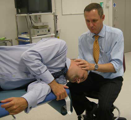
If the patient has positional vertigo (i.e. it is induced by a change in the patient's head position) then a Hallpike test should be performed (see box 1).
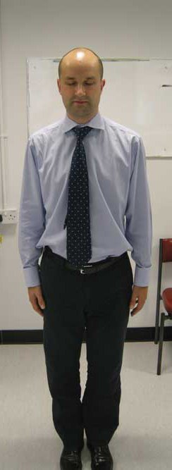
Romberg's test (figure 2) is described as positive if there is increased body sway when the eyes are closed with the patient standing with the feet close together.
The removal of visual sensory information highlights deficiencies in the other sensory functions of proprioception and/or vestibular dysfunction.
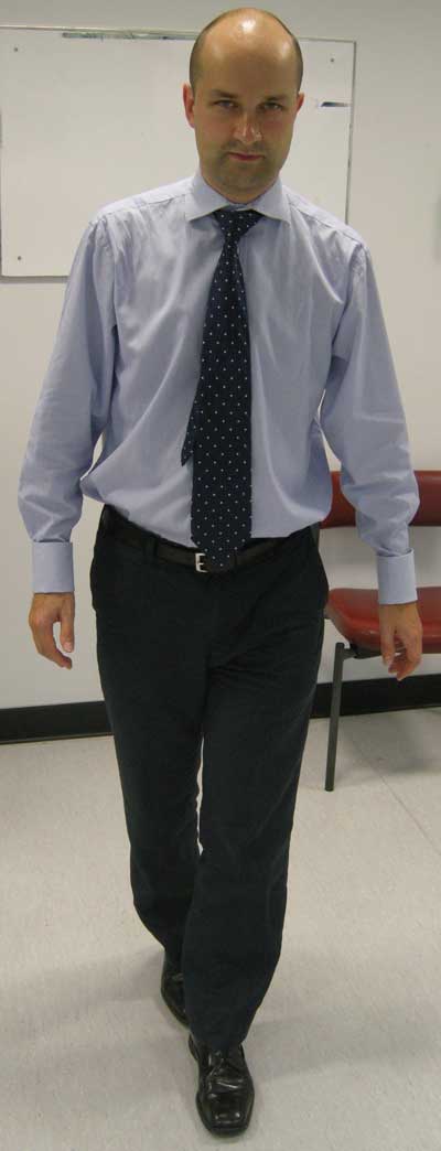
Heel to toe walking is useful to detect subtle cerebellar dysfunction, e.g. multiple sclerosis or cerebellar tumour (see figure 3).
Eyes
Examination of the cranial nerves should start with observation, in particular looking for a ptosis and any pupillary aniscoria for a Horner's syndrome from a vascular dissection or subtle IIIrd cranial nerve palsy, a false localising sign from hydrocephalus or brainstem pathology.
Visual acuity should be checked as vision is a crucial part of the balance system. The rest of the cranial nerves should be tested in particular eye movements for any ophthalmoplegia pointing to focal cranial nerve pathology and for nystagmus.
Nystagmus: In acute vertigo, due to a problem in the peripheral vestibular system (which includes the inner ear, vestibular nerves and nuclei) the slow phase of nystagmus is toward the lesion and fast phase away (contralateral) while for central causes the nystagmus is ipsilateral. Central causes tend to produce large amplitude nystagmus whereas this may only be seen in the first few days of vestibular neuritis or acute episodes of Ménières disease.
In addition it must be remembered that in peripheral causes of nystagmus visual fixation will conceal the movements and often it is not until fundoscopy is performed that the visual fixation is removed and nystagmus revealed. It should be noted that the optic disc being on the posterior pole of the eyeball will move in the opposite direction to normally observed nystagmus.
Hearing
It is generally not useful to perform clinical tests of hearing although audiometry, if available, will be helpful and is routinely performed in patients referred to an ENT clinic. Audiometry gives an objective measurement of hearing that can guide investigations and indicate the possible cause. For example, a unilateral sensorineural hearing loss will necessitate an MRI scan to exclude an acoustic neuroma.
The rest of the neurological examination should exclude evidence of central disease in particular cerebellar disease and evidence of neuropathy with wasting, weakness, absent reflexes with reduced sensation such as proprioception, vibration, pin-prick and temperature.
Systemic examination
If syncope is suspected it is wise to perform an extensive systemic examination in particular lying and standing blood pressure, and cardiovascular and respiratory system assessments.
Neurological causes
Migraine
Migrainous vertigo is often unrecognised. Criteria for diagnosis have been developed by Neuhauser.5 Definite migrainous vertigo has the four following features:
- episodic vestibular symptoms of moderate severity
- migraine according to the International Headache Criteria
- one of more of the following during two vertigo attacks - migrainous headache, headache, photophobia, phonophobia, migraine aura
- exclusion of other diagnoses
Other neurological causes include multiple sclerosis, cerebrovascular disease, and CNS tumours (e.g. acoustic neuroma).
ENT causes
Benign paroxysmal positional vertigo
BPPV is thought to arise from the presence of dense particles, most likely otoconial debris, in the posterior semi-circular canals (although the other canals can be involved). It is characterised by severe, brief episodes of rotatory vertigo provoked by changes in head position, often when the patient turns in bed or looks up. It is usually idiopathic although it can be associated with head injury or other inner ear pathology.
If Hallpike's test is positive then an Epley manoeuvre (see figure 1, and box 2) should be performed.6 This leads to resolution in up to 95% of cases.
Ménières disease
Ménières disease results from abnormal homoeostasis of the inner ear. It can begin at any age although typically starts between the ages of 20 and 40. The diagnosis is clinical and although tests have been devised to look at the electrical activity in the cochlea, they are not reliable and their place in diagnosis is somewhat controversial.
The typical symptom complex consists of episodic rotatory vertigo associated with reversible hearing loss and tinnitus. Patients often describe fullness or tinnitus occurring before the onset of vertigo. The vertigo lasts for less than 24 hours. Symptoms may not be typical however and with time patients often develop persistent sensorineural hearing loss and a sense of instability which may be punctuated with episodic rotatory vertigo.
Ménières disease is relatively uncommon and tends to be overdiagnosed in general practice.7 Treatment consists of a low-salt diet, betahistine (thought to increase cochlear blood supply) and sometimes diuretics. In the past few years intratympanic injection of gentamicin has been shown to be effective although it carries a risk of hearing loss. Vestibular nerve section and labyrinthectomy are only occasionally performed.
Labyrinthitis (vestibular neuronitis)
Labyrinthitis is a cause of acute vestibular failure and may follow a URTI. Labyrinthitis should only be diagnosed when there is associated hearing loss, as the cochlea is part of the labyrinth. There is some evidence that it is the vestibular nerve ganglia that are affected by Herpes simplex infection causing vestibular neuronitis.8, 9 Labyrinthitis can be caused by bacterial infection as a complication of otitis media.
Acute vestibular failure
Acute vestibular failure presents with acute rotatory vertigo with associated nausea and vomiting. This can be an extremely unpleasant experience for the patient who is often initially confined to bed. Unlike the majority of patients with vertigo, they will have nystagmus.
Treatment is supportive with medication to suppress vestibular activity such as prochlorperazine which may be given sublingually, im or by suppository. The symptoms gradually improve as the brain compensates for the disparity of vestibular input from the two sides. By six weeks the symptoms have more or less resolved although some patients fail to compensate and will go on to suffer chronic vertigo. We are unsure why this is, but psychological factors may be important in some patients.4
Acute vestibular failure can also be caused by head injury, with or without temporal bone fracture, drug treatment (e.g. aminoglycosides) and surgery (as a complication of mastoid surgery).
Other ENT causes
These include:
- labyrinthine fistula (generally caused by cholesteatoma)
- perilymph fistula (leakage of perilymph usually due to some form of trauma)
- autoimmune disease of the temporal bone
Cervical vertigo
There is some evidence that vertigo can be caused by arthritis affecting the high number of proprioceptive fibres originating in the neck. The condition is however ill defined but should be considered.
The elderly
The cause of vertigo in the elderly is often multifactorial. Decompensation from previous vestibular failure can occur through impaired vision and proprioception, and side effects from medication should also be considered. As with all patients with vertigo, a careful history will often point to the diagnosis. A common mistake is to prescribe vestibular sedatives such as prochlorperazine as they reduce vestibular input to the brain which in turn impairs rehabilitation.
Management
It may be of some comfort to GPs to know that a definitive diagnosis is not made in a significant number of vertiginous patients referred to a specialist, even after investigation.
If a specific disorder is identified then this can be managed with the appropriate treatment, e.g. the Epley re-positioning manoeuvre in BPPV or betahistine in Ménières disease.
Patients with migraine should be given lifestyle advice and, if required, migraine prophylaxis such as beta blockers.
Non-disease specific vestibular suppressants may be used for short-term symptomatic control but should be avoided in the long term as they prevent the process of compensation and are frequently a contributing factor to long-term dizziness.
Vestibular rehabilitation (see box 3) is usually reserved for chronic cases of dizziness as the majority of patients spontaneously compensate. Patients with chronic dizziness benefit greatly from directed exercises and support in overcoming apprehension, challenging symptoms with head movement and postural exercises.
The aim of the rehabilitation is to maximise the natural ability of the CNS to compensate for lesions in the vestibular system.
Patient education
Vertigo and dysequilibrium are particularly disturbing experiences and often lead to anxiety and loss of confidence compounding the vestibular dysfunction. Information may be obtained from the Vestibular Disorders Association, see Useful information box, opposite.
Referral
When to refer will depend to some extent on the experience of the individual GP and their access to investigations.
If a central cause is suspected or the vertigo is not being controlled then the patient should be referred. Difficulty in visualising the patient's tympanic membranes can also be a problem, e.g. because of wax, which may necessitate referral.
Often referral for vestibular rehabilitation is not accepted direct from GPs and patients will have to go through ENT or neurology. Some audiology departments have a protocol for direct referral although this is by no means universal.
We would recommend that GPs learn to perform the Hallpike test and the Epley manoeuvre as most patients will respond well and not require onward referral.
Conclusion
The dizzy patient covers a wide range of potential diagnoses. A systematic approach will lead to successful management of this initially daunting task.
REFERENCES
1 Yardley L, Owen N, Nazareth I et al. Prevalence and presentation of dizziness in a general practice community sample of working age people. Br J Gen Pract 1998;8:1131-5
2 McCormick A, Fleming D, Charlton J. Morbidity statistics from general practice: fourth national study, 1991-1992, Office of Population Censuses and Surveys. London.1995
3 Gliddon CM, Darlington CL, Smith PF. GABAergic systems in the vestibular nucleus and their contribution to vestibular compensation. Prog Neurobiol 2005;75:53-81
4 Luxon LM. Evaluation and management of the dizzy patient. J Neurol Neurosurg Psychiatry 2004;75 (Suppl IV):iv45-iv52
5 Neuhauser H, Leopold M, Von Brevern M et al. The interrelations of migraine, vertigo, and migrainous vertigo. Neurology 2001:56:436-41
6 Epley J M. The canalith repositioning procedure: For treatment of benign paroxysmal positional vertigo. Head and Neck Surgery 1992;107:399-404
7 Wladislavosky-Waserman P, Facer GW, Mokri B et al. Menieres disease: a 30 year epidemiological and clinical study in Rochester Mn. Laryngoscope 1984;94:1098-10
8 Hotson JR, Baloh RW. Acute Vestibular Syndrome. N Engl J Med 1998;339:680-685
9 Furuta Y, Takasu T, Fukuda Y et al. Latent Herpes simplex virus type in human vestibular ganglia. Acta Oto-Laryngologica 1993;113:85-89
USEFUL INFORMATION
The Vestibular Disorders Association provides support and advice for patients and healthcare professionals
ARTICLE IN PDF
 Download this article in PDF format