SEARCH

Practitioner 2010; 254 (1732): 15-18
History central to diagnosing myasthenia gravis
21 Sep 2010
AUTHORS
Dr Jennifer Spillane MB BCh BAO BMedSc MRCPI
John Newsom-Davis Fellow
Professor Dimitri Kullmann MA DPhil FRCP FMedSci
Professor of Neurology
UCL Institute of Neurology, Queen Square, London
Article
Myasthenia gravis (MG) is aptly named. It manifests as grave muscle weakness that can significantly impact on quality of life and is sometimes fatal. It is caused by failure of chemical transmission at the neuromuscular junction.
MG has a prevalence of around 20 per 100,000, so an average practice may expect to have only one patient.1 It is important not to miss the diagnosis because the condition is eminently treatable. The incidence is bimodal with a 2:1 female to male ratio in the younger population and a reversed sex ratio over the age of 60. The reported incidence has been increasing since the mid-1980s, mainly because of increased recognition of late-onset disease.2,3
However, the condition is probably still underrecognised in the very elderly and may be mistaken for other disorders such as stroke, motor neurone disease or Parkinson's disease.
Pathogenesis
MG is an autoimmune disorder in which antibodies interfere with neuromuscular transmission.4 Circulating antibodies to acetylcholine receptors (AChRs) can be detected in approximately 85% of patients.
In the remaining cases, some have antibodies that recognise muscle-specific kinase (MuSK), which is involved in the clustering of AChRs at the neuromuscular junction, and a small number of patients are persistently seronegative.5,6
In addition, very rare congenital myasthenic syndromes are recognised that present early in life. They are caused by inherited mutations affecting a variety of neuromuscular junction proteins, including enzymes involved in ACh synthesis or its breakdown following release, AChRs, and associated proteins that are necessary for the clustering of AChRs.
The thymus has an important role in the pathogenesis of MG. Approximately 15% of cases are associated with a thymoma. In non-thymomatous MG, especially in young patients, the gland is frequently enlarged with lymphocytic infiltrates. The thymus is more frequently atrophic in patients with late-onset MG, and its pathogenic role in this situation is less clear.
Presentation and clinical course
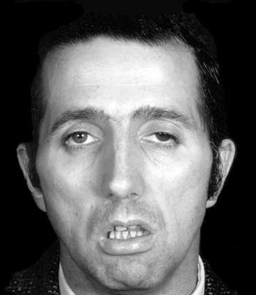
MG presents with fatiguable painless muscle weakness. Diplopia and ptosis are the most common initial presenting features. Approximately 80% of patients presenting with ocular MG will subsequently develop more generalised weakness. However, if the symptoms remain purely ocular for more than two years, the chance of progression falls to 10%.7 Bulbar and facial weakness cause difficulties with speech, chewing and swallowing, and increase the risk of aspiration. Neck weakness can lead to head drop and neck pain. Limb weakness is often proximal. Respiratory muscle weakness, the most serious manifestation of MG, can be fatal.
The mortality rate in MG has dropped markedly from >30% in the 1940s to 8MuSK MG has a relatively greater predilection for bulbar and facial muscles, with a marked female preponderance and a younger age at disease onset. MuSK MG is typically more refractory to treatment and more aggressive immunotherapy may be required.1, 2 Patients who are negative for both AChR and MuSK antibodies tend to resemble AChR antibody positive patients in clinical presentation and thymic pathology.3
Diagnosis
A detailed history is the most valuable tool in the diagnosis of MG (see table 1). The history should elicit the pattern of weakness, severity and diurnal variation. Exacerbating factors including extremes of weather, emotional stress, menstruation and intercurrent illness should be enquired about. Patients with autoimmune MG are more likely to have additional organ-specific autoimmune diseases.
If a diagnosis of MG is suspected on clinical grounds, the patient should be referred to a neurologist as certain serological tests (e.g. MUSK antibodies) and specific electrophysiological tests for MG are only carried out in specialist centres. Immunosuppressive therapy should only be initiated in a specialist setting.
There are a number of diagnostic tests (see table 2). No one test is 100% sensitive and patients who have negative antibodies and normal neurophysiology may still have MG.
Antibody tests may be repeated as a seroconversion rate of up to 15% has been reported. Some patients may only have antibodies detected on a very specific cell-based assay. In a small number of patients, the diagnosis may be based on meticulous history taking and clinical examination.
Treatment
MG treatment should be directed at ameliorating weakness with acetylcholinesterase blockers and modulating the immune system.11 There are, however, relatively few randomised controlled trials.
Acetylcholinesterase inhibitors
Acetylcholinesterase inhibitors reduce the breakdown of ACh following its release into the cleft of the neuromuscular junction. The European Federation of Neurological Societies recommends their use as first-line treatment for all forms of MG (class IV evidence), although most patients will eventually require immunomodulatory treatment.11 Pyridostigmine is the most widely used, but its short half life means that 3-4 hourly dosing is required, with the dose titrated according to symptoms. Side effects include hypersalivation, lacrimation and diarrhoea. Cholinergic crisis can occur when high doses are used. Anticholinesterases are less effective in MuSK antibody-positive MG.
Immunomodulatory therapy
Most patients with generalised MG require immunomodulatory therapy. Prednisolone is generally used as the first-line agent, although long-term side effects limit its use. The minimum effective dose should be used with appropriate monitoring of bone density and use of bisphosphonates and other bone protection agents. Steroids are anecdotally reported to lead to a transient worsening of myasthenic symptoms, and so prednisolone should be initiated cautiously at a low dose. Patients may require hospital admission for monitoring if the degree of weakness does not allow a very slow escalation of treatment.
Azathioprine is the most widely used second-line immunosuppressive agent. Trial evidence supports its use mainly as a steroid-sparing agent, although it can take 6-24 months to exert this effect.12 Side effects include leucopaenia and liver toxicity. A thiopurine methyltransferase (TPMT) assay should be requested before commencing treatment as patients with reduced activity of this enzyme become toxic at lower doses. A shared care protocol allows appropriate monitoring of haematological and hepatic parameters.
The place of mycophenolate mofetil in MG is controversial. Two randomised controlled trials failed to demonstrate a significant benefit, but its relatively mild side effect profile continues to attract support for its use.13
Methotrexate is preferred by some neurologists, although it is contraindicated in women of child-bearing age.
Third-line immunosuppressive agents used in MG include cyclophosphamide, tacrolimus, ciclosporin and rituximab, although there is a paucity of randomised controlled trial evidence.
Thymectomy
Thymectomy is commonly performed for MG, although again the evidence supporting its use is mainly retrospective. It is indicated when a thymoma is suspected, and most neurologists would advocate its use in patients presenting with MG before the age of 50. There is no evidence that patients with MuSK MG benefit from thymectomy.
Myasthenic crisis
In spite of the availability of symptomatic and immunomodulatory treatment, up to 20% of patients will experience a myasthenic crisis requiring hospital admission for ventilatory support at some stage of the disease.14
In this situation intravenous immunoglobulin (IVIG) and plasma exchange are both rapidly effective and can bring about an improvement within a few days, which may avoid the need for endotracheal intubation.
Their efficacy is similar but IVIG is better tolerated. A small minority of patients with refractory MG require regular admission for IVIG or plasma exchange.
Although MG is eminently treatable its often unpredictable fluctuations and the fear of developing a crisis can make life difficult for patients. Support from specialist nurses and other patients is available from the Myasthenia Gravis Association, see Useful information box.
Conclusion
MG is a chronic disease and the immunosuppressive treatment that is often needed requires a long-term commitment from both the health practitioner and the patient. The GP can provide enormous support to the patient on immunosuppressants. Full blood count as well as liver and renal function must be checked on a regular basis. Steroid-related side effects must also be monitored frequently to ensure that the patient is on appropriate bone protection therapy.
The GP may be the first port of call in the case of a sudden worsening of symptoms and in such cases, should refer the patient urgently to their neurology centre for specialist treatment.
REFERENCES
1 Meriggioli MN, Sanders DB. Autoimmune myasthenia gravis: emerging clinical and biological heterogeneity. Lancet Neurol 2009; 8(5):475-490
2 Phillips LH. The epidemiology of myasthenia gravis. Ann NY Acad Sci 2003; 998:407-412
3 Casetta I, Groppo E, De Gennaro R et al. Myasthenia gravis: a changing pattern of incidence. J Neurol 2010; (in press) DOI: 10.1007/s00415-010-5651-z
4 Conti-Fine BM, Milani M, Kaminski HJ. Myasthenia gravis: past, present, and future. J Clin Invest 2006; 116(11):2843-2854
5 Deymeer F, Gungor-Tuncer O, Yilmaz V et al. Clinical comparison of anti-MuSK- vs anti-AChR-positive and seronegative myasthenia gravis. Neurology 2007; 20;68(8):609-611
6 McConville J, Farrugia ME, Beeson D et al. Detection and characterization of MuSK antibodies in seronegative myasthenia gravis. Ann Neurol 2004; 55(4):580-584
7 Grob D, Brunner N, Namba T et al. Lifetime course of myasthenia gravis. Muscle Nerve 2008; 37(2):141-149
8 Barohn RJ. Standards of measurements in myasthenia gravis. Ann N Y Acad Sci 2003; 998:432-439
9 Reddy AR, Backhouse OC. "Ice-on-eyes", a simple test for myasthenia gravis presenting with ocular symptoms. Pract Neurol 2007;7(2):109-111
10 Meriggioli MN, Sanders DB. Advances in the diagnosis of neuromuscular junction disorders. Am J Phys Med Rehabil 2005; 84(8):627-638
11 Skeie GO, Apostolski S, Evoli A et al. Guidelines for treatment of autoimmune neuromuscular transmission disorders. Eur J Neurol 2010; 17(7):893-902
12 Díaz-Manera J, Rojas-García R, Illa I. Treatment strategies for myasthenia gravis. Expert Opin Pharmacother 2009;10(8):1329-1342
13 Hehir MK, Burns TM, Alpers J et al. Mycophenolate mofetil in AChR-antibody-positive myasthenia gravis: outcomes in 102 patients. Muscle Nerve 2010; 41(5):593-598
14 Jani-Acsadi A, Lisak RP. Myasthenic crisis: guidelines for prevention and treatment. J Neurol Sci 2007;261 (1-2): 127-133
USEFUL INFORMATION
The Myasthenia Gravis Association provides support and advice for patients and their families
ARTICLE IN PDF
 Download this article in PDF format