SEARCH

Jareonsettasin P, Kullmann D. Diagnosis and management of epilepsy in adults. Practitioner September 2022;266(1861):11-15
Diagnosis and management of epilepsy in adults
26 Sep 2022
AUTHORS
Dr Prem Jareonsettasin MA MBBChir MRCP, Clinical Research Fellow
Professor Dimitri Kullmann MA DPhil FRCP FMedSci FRS, Professor of Neurology
UCL Queen Square Institute of Neurology, London, UK
Competing interests: None
Article
Epilepsy is the propensity of a person to have unprovoked epileptic seizures because of an underlying neurological or somatic condition. Although the prevalence is highest during adolescence and early adulthood, the incidence is highest and rising in adults over 65. According to the International League Against Epilepsy, epilepsy can be diagnosed if any of the following criteria are met: at least two unprovoked seizures occurring on separate days (seizures within 24 hours count as one event); one unprovoked seizure with at least a 60% risk of recurrence over the next ten years on the basis of associated clinical factors (such as a recent stroke or brain tumour); diagnosis of a specific epilepsy syndrome. For each paroxysmal episode it is important to establish the sequence and progression of events, whether consciousness was impaired, and how much control the patient felt over the events. Convulsive events should be described rather than given a label. Witnesses may recall automatisms (e.g. orofacial or manual) and asymmetric movements or posture (e.g. head turning or unilateral limb dystonia). Preceding symptoms/aura may be reported by the patient or a witness. The EEG can establish the diagnosis of epilepsy and distinguish between focal and primary generalised epilepsy, but a normal EEG between seizures is not uncommon in clinical practice and does not exclude a diagnosis of epilepsy. An MRI brain scan is usually mandatory. This may reveal lesions that may have triggered seizures, such as brain tumours and vascular malformations, and may also point to certain characteristic patterns that can refine the diagnosis and guide treatment. Treatment should ideally be started and managed by tertiary care epilepsy specialists.
Epilepsy is the propensity of a person to have unprovoked epileptic seizures because of an underlying neurological or somatic condition.
Epilepsy is not one disease, rather an extremely broad group of disorders with diverse clinical presentations and electrophysiological (EEG) patterns. Thus, epilepsy is highly heterogeneous with respect to aetiologies, severity, comorbidities and outcomes. Overall, there is an increase in mortality.
The incidence and prevalence of epilepsy vary globally, reflecting in large part some of the common aetiologies and socioeconomic factors. The prevalence of epilepsy in the UK is around 1 in 100.1 Although the prevalence is highest during adolescence and early adulthood, the incidence is highest and rising in adults over 65 years old. It is one of the most common neurological disorders in later life.
Several risk factors may jointly determine whether an individual develops epilepsy. In adults, important risk factors include traumatic brain injury, CNS infections, brain tumours and excessive alcohol intake. In older adults, stroke, neurodegenerative disorders (particularly Alzheimer’s disease) and brain tumours are more common causes.
Many children with severe epilepsy survive to adulthood and will transition to adult services. Although genetic causes are more common in childhood onset epilepsies, they can also present in adulthood. Malformation of cortical development can arise from inherited or somatic mutations. Several autoimmune causes of epilepsy have emerged in recent years and may respond to immunomodulation.
Diagnosis
Epilepsy is a clinical diagnosis supported by investigations, in particular EEG and neuroimaging. The diagnosis is not always evident, or may only emerge with time. Both missing a diagnosis and misattributing symptoms to epilepsy can have serious implications.
According to the International League Against Epilepsy (ILAE), epilepsy can be diagnosed if any of the following criteria are met:2
• At least two unprovoked seizures occurring on separate days (seizures within 24 hours count as one event)
• One unprovoked seizure with a risk of recurrence of at least 60% over the next ten years on the basis of associated clinical factors (such as a recent stroke or brain tumour)
• Diagnosis of a specific epilepsy syndrome
The ILAE 2017 classification of epilepsies3 defined three diagnostic levels: seizure type, epilepsy type, and epilepsy syndrome.
Key to the diagnosis is the nature of the paroxysmal events that the patient experiences. First, it is essential to establish that a paroxysmal event is an epileptic seizure as opposed to its various mimics.4 An epileptic seizure is defined as the clinical manifestation resulting from an excessive and abnormal discharge of a population of neurones. An individual may have one or many paroxysmal events – various types of seizures, non-epileptic seizures, and non-seizure events. An accurate detailed characterisation of each type of event is crucial. This may come from patient and/or witness accounts, a video brought to the consultation, or video-EEG which is the gold standard.
The following features need to be established for each paroxysmal event.
Description
It is important to establish the sequence and progression of events, whether consciousness was impaired, and how much control the patient felt over the events. Convulsive events should be described rather than given a label.
Witnesses may recall automatisms (e.g. orofacial or manual) and asymmetric movements or posture (e.g. head turning or unilateral limb dystonia).
Preceding symptoms/aura
The preceding symptoms/aura may be reported by the patient or a witness. The order of events needs to be established with the emphasis on the initial symptom. Those commonly seen in temporal lobe epilepsy include an epigastric ‘rising’ sensation, olfactory or gustatory auras, autonomic symptoms, and experiential phenomena such as déjà vu, jamais vu and fear. Conversely, symptoms of presyncope, palpitations and chest pain give clues to alternative diagnoses.
Postictal phase
The speed of recovery can be a useful pointer. It is typically slower for a seizure than for a syncopal episode.
Postictal confusion or amnesia, and evidence of tongue biting (especially lateral), incontinence and lateralising neurological symptoms or signs are suggestive of seizures.
Temporal features
The duration of the event, time of day (especially if early morning or nocturnal), and frequency should be noted.
Triggers
Potential triggers include stress, lack of sleep, light, drugs, alcohol, specific triggers (external or internal stimuli inferred from the patient’s seizure diary).
Seizure type
For each epileptic seizure the seizure type should be classified as: focal onset, generalised onset or unknown.
The diagnostic process combines the descriptive data (semiology) with information from electrophysiology and neuroimaging.
Focal seizures may occur as focal aware seizures (auras) with or without evolution to loss of awareness. The semiology depends on the anatomical localisation of the focal cerebral disease, which can affect any cortical region.5 The temporal and frontal lobes are the most common locations of focal seizures.
Any focal seizure can progress to other cortical regions and can generalise to a focal to bilateral tonic-clonic seizure which resembles a generalised tonic-clonic seizure.
Generalised seizures are typically characterised by impaired consciousness from the onset of the attack, with bilateral and symmetric motor manifestations. Although a common pattern is for tonic axial and limb muscle co-contraction to give way to clonic jerking movements, other phenomena exist, including atonic seizures (drop attacks).
The full spectrum of seizures is wide and beyond the scope of this article. However, the following important clinical points should be considered.
Focal unaware seizures (which arise from focal lesions in limbic structures or the neocortex) should be differentiated from typical absence seizures (which are generalised seizures due to disorder of thalamocortical networks, encountered almost exclusively in genetic generalised epilepsies), see table 1.
The label of generalised tonic-clonic seizure should not be taken at face value but should be explored for possible focal onset – by thorough event characterisation, or part of a genetic generalised epilepsy syndrome – by asking about early morning myoclonus and absence seizures.
Once the seizure types are established, the epilepsy type – focal, generalised, combined generalised and focal, and unknown epilepsy – needs to be determined. EEG and MRI are supportive investigations.
EEG
The EEG is a powerful tool to:6
• Establish the diagnosis of epilepsy
• Distinguish between focal and primary generalised epilepsy
• Identify certain characteristic ‘electroclinical’ syndromes (although this is more important in childhood-onset epilepsies)
• Identify certain alternative diagnoses such as metabolic and degenerative causes of disturbed consciousness, abnormal behaviour and movements such as myoclonic jerks
Especially valuable is an EEG during a seizure, with focal seizures characteristically associated with unilateral discharges at onset, while bilaterally synchronous discharges arising from a normal background rhythm point to primary generalised epilepsy. In most cases, however, the seizure frequency is too low to capture an event during a typical EEG recording session. The sensitivity can be increased with sleep deprivation or by recording continuously with a portable device for one or more days (ambulatory EEG).
A normal EEG between seizures is not uncommon in clinical practice and does not exclude a diagnosis of epilepsy. However, interictal spikes are often seen in focal epilepsy, and some other patterns such as bilaterally synchronous spike and wave discharges or occipital spikes can occasionally be elicited with provoking measures such as hyperventilation or flashing lights. These patterns typically are not sufficient to make a diagnosis but can suggest a propensity to seizures.
Neuroimaging
An MRI brain scan is usually mandatory. This may reveal lesions that may have triggered seizures and that require further investigation or treatment, such as brain tumours and vascular malformations.
It may also point to certain characteristic patterns that, if concordant with the seizure semiology and localisation, refine the diagnosis and may guide treatment. These include mesial temporal sclerosis accompanying temporal lobe epilepsy, focal abnormalities of gyri or grey-white matter boundaries suggestive of malformations of cortical development, and bilateral hippocampal abnormalities that raise the possibility of autoimmune limbic encephalitis.7
Underlying aetiologies
Underlying aetiologies – genetic, structural, infectious, metabolic, immune, and unknown – should be investigated.
The final level of diagnosis is the epilepsy syndrome: a characteristic cluster of electroclinical features often supported by specific aetiologic findings, with prognostic and treatment implications.
Comorbidities
The presence of comorbidities is integral to the diagnosis as epilepsy is a manifestation of an underlying brain problem, exclusive to the brain or part of a systemic pathology.8 At the very least, cognitive impairment and psychiatric problems should be looked for and may only emerge over time.
Remission
An individual with epilepsy can be thought of as having a lower than normal seizure threshold. Although it is often a lifelong diagnosis, it can remit.
The current ILAE definition recognises that individuals can consider themselves free from epilepsy if seizure-free and past the age of an established childhood epilepsy syndrome. Alternatively, epilepsy can be considered to have entered remission if patients have been seizure-free for at least ten years and off all antiseizure medications (ASMs) for the past five years.
Management
Pharmacological treatment
Treatment should ideally be started and managed by tertiary care epilepsy specialists. A detailed discussion of ASM regimens is beyond the scope of this article. Treatment should be tailored to the patient’s requirements - balancing the physical, psychological, and social benefits of lower risk of seizure recurrence, against drawbacks of possible drug side effects, cost, inconvenience, and psychosocial effects. A clear discussion about treatment goals is vital. Although treatment will have been initiated in an epilepsy clinic, the GP has an important role in clarifying expectations on likely outcomes, risk and logistics of therapy.
It is important to give clear instructions when to titrate drug dosage, and when to seek immediate medical attention if signs of hypersensitivity or other idiosyncratic drug reactions develop. It is important to consider the accessibility, logistics and likely adherence to the prescribed ASMs for each individual patient.
Overall, 60-70% of patients will respond to monotherapy at the appropriate maintenance dose.
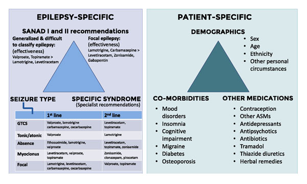
The clinician should be aware of multiple epilepsy- and patient-specific factors influencing choice of ASM (see figure 1).
Epilepsy-specific factors
Arguably the most important guidance on the choice of ASMs comes from two clinical trials that compared standard and new antiepileptic drugs (SANAD I and II) in newly diagnosed patients.9,10,11,12
In generalised and difficult to classify epilepsy, SANAD I showed that: valproate is better tolerated than topiramate, and comparable with lamotrigine; valproate is more effective than lamotrigine, and comparable with topiramate. SANAD II showed that valproate is more effective than levetiracetam.
In focal epilepsy, SANAD I found that: lamotrigine is better tolerated than carbamazepine, gabapentin, and topiramate. Carbamazepine is better than gabapentin, and lamotrigine is not inferior to carbamazepine. SANAD II showed that lamotrigine is more effective than zonisamide and levetiracetam.
From the perspective of seizure types, certain classes of drugs work better for specific types.13 First-line therapies for generalised tonic-clonic seizures are drugs acting on sodium channels (carbamazepine, phenytoin, lamotrigine) and valproate. Second-line options are levetiracetam and topiramate.
For absence seizures and myoclonus, sodium channel-targeting drugs should be avoided, as should drugs that elevate extracellular GABA levels, because they can aggravate these seizure types. Exceptions exist, and patients can have multiple seizure types concomitantly.14
Patient-specific factors
Gender
The potential teratogenicity of medications needs to be taken into account in women of childbearing age.15,16 The three main drugs considered to be safest are lamotrigine, levetiracetam and oxcarbazepine with a 2-3% risk of major congenital malformations.
Valproate should be avoided because of its very high teratogenicity (congenital malformations, reduction in IQ and neurodevelopmental disorder), and women of childbearing age should only be prescribed valproate if they are part of the Valproate Pregnancy Prevention Programme.
Topiramate and zonisamide can increase renal tract abnormalities.
For certain ASMs that have a high milk to plasma ratio (e.g. zonisamide, high-dose lamotrigine, high-dose levetiracetam), breast-fed infants should be monitored for failure to thrive.17
Age
Older adults may require lower maintenance doses of renally excreted drugs. There is also a higher risk of fractures and cardiovascular disease in older patients, therefore modifiable risk factors should be optimised.
Cognitive side effects of certain ASMs may also exacerbate existing cognitive impairment.18
Ethnicity
Patients of Han Chinese and South-East Asian ethnicity have a higher risk of hypersensitivity reactions to carbamazepine and its derivatives because of the high prevalence of the HLA-B*1502 allele.
Comorbidities
Certain ASMs can worsen or precipitate mood disturbances: levetiracetam can cause irritability; zonisamide can increase anxiety; and perampanel has been associated with suicidal and homicidal thoughts. Other ASMs such as lamotrigine and valproate have been used as mood stabilisers; pregabalin, valproate, carbamazepine and oxcarbazepine have some anxiolytic effects.
Insomnia can worsen with lamotrigine (patients may find it helpful to take the evening dose earlier). Cognitive difficulties can occur with any ASM at high dose or ASM polypharmacy but is more common with topiramate.
Valproate, topiramate and zonisamide are useful in patients with migraine. In patients with diabetes topiramate and zonisamide improve glycaemic control directly by sensitising insulin-dependent glucose uptake and indirectly through a weight loss effect.
The risk of osteoporosis is increased with enzyme inducers (e.g. carbamazepine, oxcarbazepine, phenobarbital, topiramate) and valproate. NICE recommends calcium and vitamin D supplementation and checking vitamin D every 2-5 years aiming for levels > 50 nmol/L.
Drug interactions
Enzyme inducers reduce levels of oestradiol, thus reducing the effectiveness of the oral contraceptive pill. The dosage of oestradiol should be at least 50 µg for patients with epilepsy. Lamotrigine is sensitive to progestogens. The progesterone-only pill (POP) reduces levels of lamotrigine (increasing risk of seizures), and there is also increased lamotrigine toxicity in the week off the POP. In patients established on lamotrigine, a plasma level should be measured before and after starting the POP to guide lamotrigine dosage, and patients should be advised to take the POP continuously.
Drugs that may alter metabolism or protein binding of ASMs include other ASMs, antibiotics, antidepressants, antipsychotics, and chemotherapy. Many drugs can lower seizure threshold especially tramadol, antipsychotics, tricyclic antidepressants, levodopa, thiazide diuretics, some herbal remedies, and certain antibiotics.
Non-pharmacological treatments
It is important to recognise drug resistant epilepsy, which is defined as ‘failure of adequate trials of two tolerated and appropriately chosen and used ASMs (whether as monotherapy or in combination) to achieve sustained seizure freedom’.
Any patient with drug resistant epilepsy, with or without MRI brain abnormalities, should be referred to a specialist for consideration of surgery irrespective of learning disability, psychiatric history or underlying genetic abnormalities.
Resective epilepsy surgery is the most clinically effective treatment in a subset of patients, in particular unilateral mesial temporal sclerosis, where the risk of postoperative neurological deficit is low.19
Other current treatment options include dietary therapy (e.g. a ketogenic diet) and vagus nerve stimulation.14 Various trials of novel treatment strategies (e.g. gene therapy) are ongoing or planned.20,21
Monitoring and follow-up
GPs are in a prime position to improve outcomes in a number of ways.
• Refining the diagnosis: details of new paroxysmal events of a different nature, a change in existing events, comorbidities and background information should be passed to the neurologist, who may use this to reformulate or guide further investigations
• Identifying and treating comorbidities, especially psychiatric support. The increased mortality in people with epilepsy is mainly attributable to associated comorbidities rather than epilepsy-related events
• Checking adherence to ASMs
• Monitoring effectiveness of current ASMs – frequency and duration of the patient’s stereotypical events; in addition for focal epilepsy, the frequency of secondary generalised events (focal to bilateral tonic-clonic seizures)
• Monitoring tolerability of current ASMs
• Early referral or communication with the neurologist about treatment failure and drug resistant epilepsy
• Checking and reminding patients, family/friends and carers of safety advice – especially for patients not to take baths. The most common cause of epilepsy-related deaths is drowning and sudden unexpected death in epilepsy (SUDEP)
• Signposting to patient educational resources (see Useful information box)
REFERENCES
2 Fisher RS, Acevedo C, Arzimanoglou A et al. ILAE official report: a practical clinical definition of epilepsy. Epilepsia 2014;55(4):475-82
3 Scheffer IE, Berkovic S, Capovilla G et al. ILAE classification of the epilepsies: position paper of the ILAE Commission for Classification and Terminology. Epilepsia 2017;58(4):512-21
4 Mantoan L, Kullmann DM. Evaluating first seizures in adults in primary care. Practitioner 2011;255(1743):25-28
5 Chowdhury FA, Silva R, Whatley B, Walker MC. Localisation in focal epilepsy: a practical guide. Pract Neurol 2021;21(6):481-91
6 Fowle AJ, Binnie CD. Uses and abuses of the EEG in epilepsy. Epilepsia 2000;41 Suppl 3:S10-18
7 Sidhu MK, Duncan JS, Sander JW. Neuroimaging in epilepsy. Curr Opin Neurol 2018;31(4):371-78
8 Keezer MR, Sisodiya SM, Sander JW. Comorbidities of epilepsy: current concepts and future perspectives. Lancet Neurol 2016;15(1):106-15
9 Marson AG, Al-Kharusi AM, Alwaidh M et al. The SANAD study of effectiveness of carbamazepine, gabapentin, lamotrigine, oxcarbazepine, or topiramate for treatment of partial epilepsy: an unblinded randomised controlled trial. Lancet 2007;369(9566): 1000-15
10 Marson AG, Al-Kharusi AM, Alwaidh M et al. The SANAD study of effectiveness of valproate, lamotrigine, or topiramate for generalised and unclassifiable epilepsy: an unblinded randomised controlled trial. Lancet 2007;369(9566):1016-26
11 Marson A, Burnside G, Appleton R et al. The SANAD II study of the effectiveness and cost-effectiveness of levetiracetam, zonisamide, or lamotrigine for newly diagnosed focal epilepsy: an open-label, non-inferiority, multicentre, phase 4, randomised controlled trial. Lancet 2021;397(10282):1363-74
12 Marson A, Burnside G, Appleton R et al. The SANAD II study of the effectiveness and cost-effectiveness of valproate versus levetiracetam for newly diagnosed generalised and unclassifiable epilepsy: an open-label, non-inferiority, multicentre, phase 4, randomised controlled trial. Lancet 2021;397(10282):1375-86
13 Rogawski MA, Löscher W, Rho JM. Mechanisms of action of antiseizure drugs and the ketogenic diet. Cold Spring Harb Perspect Med 2016 May;6(5)
14 National Institute for Health and Care Excellence. NG217. Epilepsies in children, young people and adults: diagnosis and management. NICE. London. 2022 www.nice.org.uk/guidance/ng127
15 Tomson T, Battino D. Teratogenic effects of antiepileptic drugs. Lancet Neurol 2012;11(9):803-13
16 Stephen LJ, Harden C, Tomson T, Brodie MJ. Management of epilepsy in women. Lancet Neurol 2019;18(5):481-91
17 Arfman IJ, Wammes-van der Heijden EA, Ter Horst PGJ et al. Therapeutic drug monitoring of antiepileptic drugs in women with epilepsy before, during, and after pregnancy. Clin Pharmacokinet 2020;59(4):427-45
18 Sen A, Jette N, Husain M, Sander JW. Epilepsy in older people. Lancet 2020;395(10225):735-48
19 de Tisi J, Bell GS, Peacock JL et al. The long-term outcome of adult epilepsy surgery, patterns of seizure remission, and relapse: a cohort study. Lancet 2011;378(9800):1388-95
20 Walker MC, Kullmann DM. Optogenetic and chemogenetic therapies for epilepsy. Neuropharmacology 2020;168:107751
21 Kullmann DM, Schorge S, Walker MC, Wykes RC. Gene therapy in epilepsy - is it time for clinical trials? Nat Rev Neurol 2014;10(5):300-04
ARTICLE IN PDF
 Download this article in PDF format
PRESET SEARCHES
PubMed
Epilepsy in primary care