SEARCH

Elliott E, Davenport RJ. History and examination pivotal in diagnosis of Bell’s palsy. Practitioner September 2021;265(1851):17-20
History and examination pivotal in diagnosis of Bell’s palsy
24 Sep 2021
AUTHORS
Dr Elizabeth Elliott BSc MB BS, Clinical Fellow in Regenerative Neurology, Centre for Clinical Brain Sciences, University of Edinburgh, Edinburgh, UK
Dr Richard J Davenport DM FRCP (Ed), Consultant Neurologist, Department of Clinical Neurosciences, Royal Hospital for Children and Young People, and Clinical Neurosciences, Royal Infirmary of Edinburgh, Edinburgh, UK
Competing interests: None
Article
Bell’s palsy is the most common cause of facial palsy and presents as a unilateral lower motor neurone facial weakness in association with sensory and parasympathetic dysfunction.1 The annual incidence of Bell’s palsy is around 38 per 100,000 in the UK.2,3
The Scottish anatomist Sir Charles Bell first demonstrated in animal dissection studies during the 1800s that cutting the seventh cranial nerve resulted in an ipsilateral facial palsy.4
Although the severity of facial weakness in Bell’s palsy varies, maximal facial weakness occurs within 72 hours and most cases recover over the following few weeks.5,6
Incomplete recovery, although uncommon, can result in permanent facial paralysis or abnormal reinnervation with considerable functional and aesthetic sequelae requiring long-term multidisciplinary team (MDT) support.
Bell’s palsy is still considered to be idiopathic, in contrast to other facial nerve palsies with known infective, malignant or autoimmune causes. Most cases of Bell’s palsy can be diagnosed and managed in primary care.
Risk factors
Bell’s palsy affects men and women equally and the incidence increases with age particularly around midlife.6 The frequency of left- and right-sided weakness is similar.7 Risk factors include: pregnancy, acute respiratory infection, immunocompromise, diabetes mellitus and hypertension.3,8 Bell’s palsy exhibits seasonal variation, increasing in colder months around the world.7,9,10
Risk factor reporting varies across studies, adverse prognostic factors include: decreased lacrimation, complete paralysis, advanced age, hypertension, diabetes mellitus and pain in areas other than the ear (including mastoid and facial pain).3,4,7
Aetiology
The underlying pathophysiology of Bell’s palsy is not fully understood and there are several causative theories relating to the basic mechanism of facial nerve inflammation, oedema and compression. The facial nerve is particularly vulnerable to compression and symptomatic ischaemia as it passes through the temporal bone in the narrow facial canal.4,11,12
The identification of herpes viral reactivation in a proportion of cases, particularly herpes simplex virus 1 (HSV-1) and varicella zoster virus (VZV), has led to the hypothesis that these are causative agents.5,13,14 However, viral detection results vary across studies and notably in one study the presence of herpes viral DNA did not appear to influence clinical recovery outcomes.5,15
The first gene variant associated with Bell’s palsy was recently identified in a meta-analysis, however the biological mechanisms underlying the increased associated risk are yet to be defined.16
Differential diagnoses
It is essential to consider alternative causes of acute peripheral facial palsy including: infection, malignancy and autoimmune disease (see table 1).
The most common infective causes of acute peripheral facial palsy include herpes zoster (Ramsay Hunt syndrome) and Lyme neuroborreliosis.17
The diagnostic clue in Ramsay Hunt syndrome is herpetic lesions in the external ear canal, however these may appear after the facial palsy or not at all in the case of zoster sine herpete.13,17
Other distinguishing features of Ramsay Hunt syndrome include severe ear/postauricular pain and audiovestibular nerve involvement.18
Lyme neuroborreliosis should always be considered for patients in an endemic area or who have characteristic features, so it is important to ask about recent travel, tick bites, rashes etc.
Multiple cranial palsies are suggestive of sinister causes such as compressive or infiltrative neoplastic lesions.
Systemic features may suggest basal meningitis caused by infective, malignant or inflammatory diseases.17,18
Presentation
Bell’s palsy is a clinical diagnosis. Patients typically present with an acute onset (ranging from a few hours to 72 hours) unilateral lower motor facial weakness (see box 1). The degree of weakness varies significantly and may be preceded or accompanied by: postauricular pain, altered taste, dry eye and hyperlacrimation.21 These associated non-motor symptoms reflect the involvement of sensory and parasympathetic facial nerve pathways.4 In some cases, onset may be so acute as to mimic stroke. Patients often complain of sensory symptoms affecting the face, although the facial nerve is purely motor as regards the face.
A clinical diagnosis in primary care is acceptable if the above features are confirmed but it is important to identify any unusual features, including red flags which require referral for further investigation (see table 2).
Focused history
The following key areas should be addressed when taking the history:
Timing
• Was the onset more than three days ago? Bell’s palsy never presents insidiously.
• How has the weakness evolved over this time? Maximal weakness in Bell’s palsy occurs within 3 days, deviation from this course is atypical.1
The frequency of recurrence ranges from 6.5 to 19.4% and recurrent episodes are associated with lower recovery rates.22 Recurrence of symptoms within weeks or months should prompt further investigation.
Severity
• Is there incomplete eye closure?
All patients with eye closure weakness require an eye care regimen consisting of lubricating daytime eye drops with optional patching to protect against injury and eye ointment with taped closure at night. Inability to close the eye completely leaves the patient at risk of corneal ulceration and advice from an ophthalmologist may be required.19
Eating, communication and aesthetics
Patients may experience difficulty eating, drinking, or speaking.
The psychopathology associated with facial disfigurement, even temporary, should not be underestimated and reassuring patients that a good prognosis is likely is important.
Additional associated symptoms
Accompanying symptoms include: postauricular pain in around half of patients, altered taste due to chorda tympani involvement in around a third, eye dryness in around a third and dry mouth in around a fifth.
Additional symptoms may include: facial pain, hyperlacrimation, aural fullness and loud noise intolerance (hyperacusis).21
Isolated facial nerve deficits
In particular the absence of diplopia, dysphagia or any audiovestibular symptoms such as hearing loss, tinnitus, vertigo. Patients may complain of altered sensation which appears to be related to the weakness.
Systemic symptoms
Enquire about, and exclude, features of systemic disease (e.g. weight loss, fever, night sweats, general unwellness etc).
Past medical history
Malignancy, particularly cutaneous malignancy affecting the head and neck, a history of trauma, previous stroke or other vascular risk factors should be noted.
Examination
A diagnosis of Bell’s palsy can only be made in the absence of other neurological or systemic signs suggestive of an alternative cause.
General head and neck
It is important to inspect the skin for lesions, check the anterior two thirds of the tongue and soft palate for herpetic lesions, palpate to check for a parotid gland mass and enlarged lymph nodes.18 The patient should be assessed for otitis media or vesicles in the external auditory canal.
Cranial nerve examination
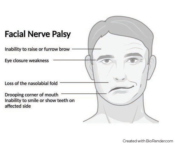
This should be carried out at rest. The patient should be assessed for loss of tone evidenced by drooping of the eyebrow, loss of forehead wrinkles, loss of the nasolabial fold, and drooping of the corner of the mouth (see figure 1).
Facial asymmetry and unilateral lower motor neurone weakness across the upper, middle and lower facial zones should be looked for. The House-Brackmann grading system has been widely used as a study outcome measure to grade facial weakness (I-normal facial function to VI-total paralysis) and is a useful reference during examination (see below).23
The House-Brackmann grading system23
I Normal symmetrical function in all areas
II Slight weakness on close inspection, able to close eye
III Obvious but not disfiguring weakness
IV Obvious disfiguring weakness and unable to close eye
V Barely perceptible movement
VI No movement
The patient should be assessed at rest and also when the face is expressive. Comparison with previous photographs may be helpful.
Assessment of facial expression
• Inability to raise or furrow the brow on the affected side (frontalis weakness). Note that forehead sparing is generally seen in upper motor neurone/central facial weakness due to the bicortical representation of the forehead. In milder cases lower facial weakness may be more apparent than forehead weakness
• Ask the patient to smile, show their teeth, whistle, puff their cheeks out. Orbicularis oris weakness causes the mouth to be drawn to the unaffected side
• Eye closure weakness. Orbicularis oculi weakness may also cause excess lacrimation as tears cannot be directed across the eye surface.19
The results of the rest of the neurological examination should be normal.
Confirming diagnosis
In most cases of new onset facial palsy no further laboratory, electrodiagnostic or imaging investigations are required although blood glucose to exclude diabetes is a reasonable precaution to take.12
Patients should be referred in the following situations:
• Urgent referral to neurology or ENT, depending on local referral pathways, if there is uncertainty about the diagnosis or there are atypical features20
In moderate to severe cases of Bell’s palsy:
• Urgent referral to an ophthalmologist if there is concern about eye safety with incomplete eye closure
• Psychological support should be considered
• Referral to a plastic surgical facial reanimation service should be considered if patients have persisting facial weakness several months following symptom onset.24
Management
Current NICE guidance recommends early treatment with a short course of high-dose prednisolone. Treatment should be started within the first 72 hr of presentation or as soon as possible within the first 2 weeks at a dose of 50 mg daily for 10 days or 60 mg daily for five days followed by a daily reduction in dose of 10 mg (for a total treatment time of 10 days).20
While corticosteroids alone have been shown to reduce the rate of incomplete recovery compared with antivirals alone, the evidence to support dual therapy, i.e. prednisolone plus an antiviral is unconvincing.25,26
The latest Cochrane meta-analysis reported, with a moderate level of confidence, that combined therapy reduced the number of long-term effects such as facial movement abnormalities compared with steroid monotherapy.27 This meta-analysis was significantly limited following the exclusion of small studies at high risk of bias.
The lack of demonstrable combined therapy efficacy may also reflect disease heterogeneity, with viral reactivation including zoster sine herpete representing only a subset of cases. Antiviral therapy does not appear to be associated with adverse effects and so it may be reasonable to treat patients empirically with the features of zoster sine herpete such as more severe facial weakness and pain with dual therapy.18,26
Surgical decompression of the facial nerve as an alternative acute intervention is not recommended.12
Between 20 to 30% of patients are left with residual weakness and in around half of these cases the deficit is moderate to severe.24,28 Aberrant reinnervation may occur 5 months or more after symptom onset resulting in motor or autonomic synkinesis, for example jaw-winking or salivation induced facial sweating.4,19 Chronic facial muscle weakness can result in the development of spasm and contractures and long-term disability.
MDT management involves a range of interventions including physiotherapy, which although not clearly evidence based, may be beneficial for patients with long-term complications.12,19 Chemodenervation using botulinum toxin can be used for troublesome aberrant reinnervation.19,29 The role of nerve grafting procedures remains uncertain.24
Conclusions
Bell’s palsy is a clinical diagnosis and most cases can be managed with acute high dose steroid therapy, an eye care regimen and reassurance in the primary care setting. A short course of oral steroids improves recovery rates. Dual steroid and antiviral therapy may be beneficial however it is not possible to draw firm conclusions from the current evidence base.
The majority of patients will recover completely within a few weeks. However, complicated cases of Bell’s palsy may need early specialist referral for MDT management.
Recurrence may occur in up to 20% of cases and should be managed in the same way as the primary episode.
REFERENCES
1 Hohman MH, Hadlock TA. Etiology, diagnosis, and management of facial palsy: 2000 patients at a facial nerve center. Laryngoscope 2014;124(7):E283-E293
2 Morales DR, Donnan PT, Daly F et al. Impact of clinical trial findings on Bell’s palsy management in general practice in the UK 2001–2012: interrupted time series regression analysis. BMJ Open 2013;3(7):e003121
3 Wijnans L, Dodd C, Weibel D, Sturkenboom M. Bell’s palsy and influenza (H1N1) pdm09 containing vaccines: a self-controlled case series. PloS One 2017;12(5), e0175539–e0175539
4 Reich SG. Bell's palsy. Continuum 2017;23(2):447-66
5 Ordoñez G, Vales O, Pineda B et al. The presence of herpes simplex-1 and varicella zoster viruses is not related with clinical outcome of Bell’s palsy. Virology 2020;549:85-88
6 Rowlands S, Hooper R, Hughes R, Burney P. The epidemiology and treatment of Bell’s palsy in the UK. Eur J Neurol 2002;9(1):63-67
7 De Diego JI, Prim MP, Madero R, Gavilán J. Seasonal patterns of idiopathic facial paralysis: a 16-year study. Otolaryngol Head Neck Surg 1999;120(2):269-71
8 Peitersen E. Bell’s palsy: the spontaneous course of 2,500 peripheral facial nerve palsies of different etiologies. Acta Oto Laryngol Suppl 2002;122(549):4-30
9 Erdur H, Ernst S, Ahmadi M et al. Evidence for seasonal variation of Bell’s palsy in Germany. Neuroepidemiology 2018;51(3-4):128-30
10 Lee JS, Kim YH. Epidemiological trends of Bell’s palsy treated with steroids in Korea between 2008 and 2018. Muscle Nerve 2021;63(6):845-51
11 Takezawa K, Townsend G, Ghabriel M. The facial nerve: anatomy and associated disorders for oral health professionals. Odontology 2018;106(2):103-16
12 Baugh RF, Basura GJ, Ishii LE et al. Clinical Practice Guideline: Bell’s palsy executive summary. Otolaryngol Head Neck Surg 2013;149(5):656-63
13 Furuta Y, Ohtani F, Kawabata H et al. High prevalence of varicella-zoster virus reactivation in herpes simplex virus-seronegative patients with acute peripheral facial palsy. Clin Infect Dis 2000;30(3):529-33
14 Murakami S, Mizobuchi M, Nakashiro Y et al. Bell palsy and herpes simplex virus: identification of viral DNA in endoneurial fluid and muscle. Ann Intern Med 1996;124(1 I): 27-30
15 Stjernquist-Desatnik A, Skoog E, Aurelius E. Detection of herpes simplex and varicella-zoster viruses in patients with Bell's palsy by the polymerase chain reaction technique. Ann Otol Rhinol Laryngol 2006;115(4):306-11
16 Skuladottir AT, Bjornsdottir G, Thorleifsson G et al. A meta-analysis uncovers the first sequence variant conferring risk of Bell’s palsy. Sci Rep 2021;11(1):4188
17 Zimmermann J, Jesse S, Kassubek J et al. Differential diagnosis of peripheral facial nerve palsy: a retrospective clinical, MRI and CSF-based study. J Neurol 2019;266(10):2488-94
18 Fuller G, Morgan C. Bell’s palsy syndrome: mimics and chameleons. Pract Neurol 2016;16(6):439-44
19 Eviston TJ, Croxson GR, Kennedy PGE et al. Bell’s palsy: aetiology, clinical features and multidisciplinary care. J Neurol Neurosurg Psychiatry 2015;86(12):1356-61
20 National Institute for Health and Care Excellence. Clinical knowledge summary. Management of Bell's palsy cks.nice.org.uk/topics/bells-palsy/management/management
21 De Seta D, Mancini P, Minni A et al. Bell’s palsy: symptoms preceding and accompanying the facial paresis. Sci World J 2014;801971–801976
22 Dong SH, Jung AR, Jung J et al. Recurrent Bell’s palsy. Clin Otolaryngol 2019;44(3):305-12
23 House JW, Brackmann DE. Facial nerve grading system. Otolaryngol Head Neck Surg 1985;93:146-47
24 Glass GE, Tzafetta K. Optimising treatment of Bell’s palsy in primary care: the need for early appropriate referral. Br J Gen Pract 2014;64(629):e807–e809
25 Sullivan FM, Swan IR, Donnan PT et al. Early treatment with prednisolone or acyclovir in Bell's palsy. N Engl J Med 2007;357(16):1598-1607
26 Engström M, Berg T, Stjernquist-Desatnik A et al. Prednisolone and valaciclovir in Bell's palsy: a randomised, double-blind, placebo-controlled, multicentre trial. Lancet Neurol 2008;7(11):993-1000
27 Gagyor I, Madhok VB, Daly F, Sullivan F. Antiviral treatment for Bell’s palsy (idiopathic facial paralysis). Cochrane Database Syst Rev 2019(9), CD001869
28 Gilden DH Bell’s palsy. N Engl J Med 2004;351;(13):1323-31
29 Butler DP, Grobbelaar AO. Facial palsy: what can the multidisciplinary team do? J Multidiscip Healthcare 2017;10:377-381
ARTICLE IN PDF
 Download this article in PDF format